Iris Cysts

By Paul T. Finger, MD

Description

Cysts can form in different parts of the iris and ciliary body. Most remain undetected, unless they push on the iris or get relatively large. The most common is the neuro-epithelial iris cyst, which is typically located beneath the iris root. Cysts can also be located in the ciliary body, the iris stroma, and be formed by splitting (schisis) of the iris pigment epithelium). These cysts can push the iris forward, appear as a mass or tumor and may cause angle closure glaucoma.

Symptoms

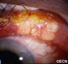
Almost all iris cysts are located behind the iris and cause no symptoms. They are found by the eye care specialist during ophthalmic examination. Iris stromal cysts can become visible on the surface of the iris. Most commonly, the eye doctor sees a bulge in the iris stroma and considers that a tumor may be pushing it forward. At that point, the doctor may send the patient for an ocular tumor evaluation (to look behind the iris with ultrasound or OCT imaging).

Diagnosis

Most iris cysts can be diagnosed by clinical examination with high-frequency ultrasonography.

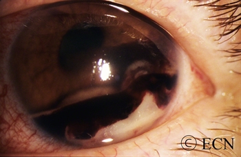
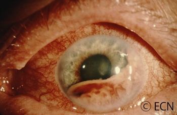
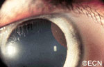
Large cysts can be seen by routine slit-lamp examination.

High frequency ultrasonography reveals internal echolucency consistent with the diagnosis of iris stromal cyst.
This iris stromal cyst extends from the iris stroma onto the corneal endothelium.


Though cysts are benign, periodic observation is necessary due to the rare instances of secondary angle closure glaucoma (only seen with giant cysts, multifocal cysts and iris schisis cysts). 

Slit-lamp photograph of an Iris Pigment Epithelial Cyst. Note its smooth, rounded surface.
High-frequency ultra-sonography of a neuro-epithelial iris cyst. Note anterior displacement of the iris with focal angle closure.


High-frequency ultrasonography (aka UBM) has revolutionized our ability to image and thus “see” tissues behind the iris. It has allowed for evaluation of tumor thickness, depth of penetration, and invasion of adjacent tissues. Ultrasounds ability to determine if a tumor is solid or cystic has been particularly helpful.

Treatments

  • Most iris cysts do not require treatment.
  • Treatment is performed for the rare instances of secondary glaucoma or when the visual axis (pupil) becomes blocked. Laser has been used to deflate iris pigment epithelial cysts with minimal side effects.
  • The natural history of iris cysts has not been determined. In my experience, some get smaller, some get larger and most stay the same. Until eye care specialists understand which cysts will grow, periodic observation is warranted.


Related links


Anterior Uveal Metastasis

By Paul T. Finger, MD

Description

Metastatic Breast Cancer

Intraocular metastasis is the most common malignant intraocular tumor and may occur in as many as 10% of patients with metastatic cancer. Most of these tumors go undetected unless they affect vision and become symptomatic.

Metastatic breast cancer is the most common in women and lung is the most common in men. Other primary sites include prostate, skin, kidney, colon and thyroid. Leukemia and lymphoma also occur in the eye.

Less than 10 percent of intraocular metastatic tumors are located in the anterior part of the eye.

Metastatic Lung Cancer

Symptoms

Most patients with iris metastasis present either with a visible cance (see photographs) or tumor-related “secondary” glaucoma. Glaucoma can cause foggy vision, halos around lights and headaches (brow aches). These “symptomatic” patients that come to or are sent for ophthalmic examination.

Most iris metastasis patients either have a history of cancer or are found to have a primary source of the tumor (on systemic examination). It is important to perform complete systemic survey when intraocular metastasis is suspected. This examination should include radiographic imaging (staging).

The New York Eye Cancer Center protocol involves whole body, scalp-to-toes PET/CT.

The reason is that though most intraocular cancers come from the lung and breast, there exist multiple less common “source” primary cancers. PET/CT evaluates the entire body. Consultation with an adult or pediatric medical oncologist should be obtained.

Despite systemic evalutions, sometimes patients will have no detectable primary cancer. Most of these tumors will later be found to originate from the lung (particularly in men). In these cases (where no primary cancer is found), the intraocular tumor becomes the only tissue that can be used to direct the search for its site of origin. Thus, biopsy of an anterior segment metastasis may become necessary. Biopsy techniques include: fine-needle aspiration biopsy, transcorneal tumor excision, and the relatively safe “Finger Iridectomy Technique (FIT).”

Treatments

Advanced anterior iris and ciliary body metastases can be difficult to manage. Radiation therapy alone or in combination with intraocular anti-VEGF drug therapy may be used to control the tumor, protect vision and conserve the eye. If left untreated, metastatic cancer in the iris can cause glaucoma and a blind painful eye. Thankfully, symptomatic anterior segment metastases are much less common than choroidal metastasis.

When the diagnosis of intraocular metastasis is made or suspected, eye cancer specialists always dilate and examine both eyes. This is because metastases can be both bilateral and multifocal. Computed tomography or magnetic resonance imaging of the brain and lungs should be performed due to a high incidence of concurrent metastases.

Related links


Intraocular Lymphoma (Vitreoretinal Lymphoma)

By Paul T. Finger, MD

Description

Intraocular Lymph
Choroidal infiltration by lymphoma

Most patients diagnosed to have intraocular lymphoma have symptoms of vitreous floaters, a history of systemic lymphoma or have been diagnosed as having chronic uveitis. Any patients with vitreous cells, no history of recent intraocular surgery and a non-painful eye should be suspected to have intraocular lymphoma.

Symptoms

Case Example: A 70-year-old woman was noted to experience an acute deterioration of her vision due to vitreous cells. A diagnostic vitrectomy was performed. Pathology showed large B-cell lymphoma. A complete metastatic survey (imaging studies of the brain, chest, abdomen), lumbar puncture and bone marrow biopsy were found to be negative.

Diagnosis

The diagnosis of intraocular lymphoma is typically made by removal of cells (vitrectomy biopsy) from the eye with subsequent cytopathologic evaluation. Lymphoma biopsy should be performed at a center with ophthalmic pathology services used to working with small specimens. Once the diagnosis is certain, a hematologist-oncologist should be consulted to perform an evaluation for systemic and central nervous system lymphoma (staging).

Treatments

Intraocular Lymphoma B Scan
B-scan ultrasound reveals an irregular retinal surface and variable internal reflectivity.

Though chemotherapy can be used to treat the systemic disease, poor intraocular drug penetration can leave residual lymphoma in the eye. In these cases, external beam radiation therapy to the eyes and orbits will typically clear the intraocular disease.

The literature consists of a multitude of case-reports and no evidence based comparative studies. In my experience, most patients are eventually treated with ocular irradiation. Others have had prophylactic whole-brain irradiation as well as combinations of local and systemic chemotherapy.

What is common to most of these reports is that, when the lymphoma is found in the brain, the prognosis is poor. Clearly, a natural history study followed by prospective randomized treatment trials would be helpful to find the cure for patients with intraocular lymphoma.

Additional info

Radiation Therapy

Radiation therapy has been the most common treatment for intraocular lymphoma. Both eyes are usually treated because approximately 80% of cases will either present as or go on to develop bilateral disease (within 8 years).

The prescription dose has been decreasing with less than 3,000 cGy being employed in many centers. The dose to the orbit is adjusted when whole-brain irradiation is required. With this in mind, remember to always obtain a complete neurological work-up to rule out central nervous system (CNS) involvement. Radiation will help acutely, but intraocular and CNS relapse are common.

Chemotherapy

Recent investigations have combined radiation therapy with systemic or intrathecal chemotherapy. Several studies suggest that this approach has prolonged survival. Chemotherapeutic agents have been given intravenously, intrathecally, and intravitreally (into the eye).

Injections of chemotherapy into the eye have been investigated as an alternative to radiation. Typically using methotrexate and/or retuximab, multiple injections are required and carry the known risks associated with ocular perforation (e.g. infection) as well as chemotherapy associated side-effects. This is why most centers use intravitreal chemotherapy when patients demonstrate intraocular recurrence after failure of radiation therapy.

Related links


Infiltrative Choroidopathy

By Paul T. Finger, MD

Description

In this case, our patient had a history of myeloma. If suspected, the work-up for multiple myeloma includes: a bone-marrow biopsy, skeletal x-rays, serum protein electrophoresis, urinalysis for Bence-Jones proteins. Several cases have described orbital involvement.

In this case, progression was documented and external beam radiation therapy stabilized her ocular condition.


Chronic Lymphocytic Leukemia

By Paul T. Finger, MD

Description

Leukemic Infiltrate of the Anterior Chamber - Hypopyon with Hyphema
Leukemic Infiltrate of the Anterior Chamber – Hypopyon with Hyphema in a 6-year old child

Leukemia is a systemic disease that can involve the eyes. It occurs in both children and adults. In that the eyes can be a relative sanctuary (a place to hide from systemic chemotherapy) for leukemic cells, intraocular recurrences have been reported after both total body irradiation and systemic chemotherapy. It is reasonable for all patients with leukemia to have periodic eye examinations.

Adults can also have leukemia and develop infiltration of the eyes. The case presented in this section (below) demonstrates one such case.

Symptoms

Depending on the location of the leukemic infiltrates, patients will have different symptoms. Most patients are found to have retinal tumors with hemorrhage, optic nerve swelling (papilledema) or cells in the anterior chamber of the eye (hypopyon/hyphema). Leukemic tumor called “chloroma” can also form around the eyes and in the orbit.

In this case, posterior uveal infiltration caused thickening of the choroid that moved the retina forward. This made the patient more far-sighted (hyperopic shift) due to a shortening of the distance between the cornea/lens complex and the retina.

The front of the eye was also infiltrated by leukemic cells. Thickening of the iris, ciliary body and anterior uvea induced a secondary “narrow angle” glaucoma. The symptoms of glaucoma include seeing haloes around lights, headache (brow-ache) and eye pain.

Diagnosis

The clinical diagnosis of infiltrative ocular leukemia (e.g. typical findings and a history of systemic leukemia) is typically adequate for treatment of patients. When proof of recurrence is needed to proceed with systemic or local therapy, a biopsy may be performed for a cytologic or histopathologic diagnosis.

Treatments

The treatment of intraocular leukemia is (in part) dependent on the treatment of the patient’s systemic disease. For example, in the clinical situation where the eye findings are not posing an immediate threat to vision, your physician my wait for systemic therapies to resolve the ocular disease. In cases where all standard treatments have been unsuccessful in resolving the ocular leukemia; patients will be treated with low-dose external beam radiation therapy.

Related links


Squamous Carcinoma of the Eyelid

By Paul T. Finger, MD


Note the relatively flat surface, red (indurated – inflammed edges, and the white flakey material on the right side of its surface.

Description

Squamous carcinomas of the eyelid can locally invade the orbit and sinuses, but rarely metastasizes. It is the second most common malignant eyelid tumor, but is 10 times less common than basal cell carcinoma. This should not be confused with conjunctival squamous carcinoma, which is the most common conjunctival cancer and can spill over onto the eyelid skin.

Symptoms

Squamous eyelid carcinoma can have symptoms that range from the appearance of a hypervascular flat pale, reddish or flaky lesion on the eyelid skin to a thickened well-demarcated reddish, flat tumor surrounded by inflammation (with or without scaling from its surface).

Diagnosis

Squamous carcinoma of the eyelid should be photographed at baseline. These lesions can remain unchanged (for years), then invade into the dermis and grow. A simple wedge biopsy can be performed and sent for pathologic evaluation. Once the diagnosis of squamous carcinoma is biopsy proven, definitive treatment is needed.

Treatments

Like basal cell carcinomas, squamous cell cancers of the eyelid rarely metastasize. However, they can grow around the eye into the orbit, sinuses and brain. Therefore, early intervention with complete excision or destruction is warranted.

Surgical approaches depend on the doctors training. Eye care specialists will either perform a planned excision with frozen-section control (of the margins) or the Moh’s Technique. Both types of surgery are used to remove the entire tumor along with a safety zone of normal appearing tissue from the edges of surgical wound (margins). No comparative studies have definitively shown that one technique better than the other. Both surgeries require a cosmetic surgical repair to return eyelid function and cosmesis. When the orbit and sinuses are not involved, local excision is usually curative.

Extension into the orbit and sinuses typically requires more extensive surgery (exenteration, sinusectomy) with subsequent radiation therapy.

At the New York Eye Cancer Center, we have treated select patients with small squamous carcinomas of the eyelid with topical chemotherapy agents or freezing (cryotherapy) using “Finger-tip” applicators. Dr. Finger tries to avoid surgery when possible.

Related links


Sebaceous Carcinoma of the Eyelid

By Paul T. Finger, MD

Description

Sebaceous carcinoma arises from the glands within the eyelids, caruncle or eyebrow. They are more commonly found on the upper eyelid and in middle-aged patients.

Symptoms

Sebaceous cell carcinoma is suspected due to evidence of eyelash loss and the formation of a yellow-nodule. This tumor can also present as a persistent (months) non-responsive blepharitis or conjunctivitis. In these cases, a high index of suspicion for sebaceous cell carcinoma will lead to biopsy and the diagnosis. Once sebaceous carcinoma is suspected a biopsy is warranted. Before surgery, the pathologist should be advised of this possible diagnosis so the specimen can be processed appropriately

Sebaceous carcinomas are one of the rarest eye cancers and can look like a chalazion (stye) or conjunctivits. In that sebaceous carcinoma can mimick these relatively benign dieases, eye care specialists should be suspicious of this tumor in any patient with persistent conjunctivitis, blepharoconjunctivitis or chronic/recurrent chalazion. Therefore, any conjunctivitis or chalazion that is not getting better after 3 months of observation, should be biopsied.

Diagnosis

Sebaceous carcinoma of the eyelid typically presents as a yellow-nodule in the upper lid. The patient may have history of colon, breast, genitourinary or other skin malignancies (Muir-Torre Syndrome).* When a nodular tumor, it can cause eyelash loss, but is not painful (compared to styes). This tumor can also present as a hypervascular conjunctival tumor and tends to spread along the conjunctival surface. In these cases sebaceous carcinoma looks like conjunctivitis.

It is worth repeating that “any conjunctivitis or chalazion that is not getting better after 3 months of observation should be biopsied.” If cytopathology is available at your medical center, a scrape biopsy of the conjunctiva can be initially performed in the office setting. However, the gold standard for diagnosis of sebaceous carcinoma is histopathologic evaluation of a fresh tissue specimen.

Once the diagnosis is made, a metastatic survey is warranted. Sebaceous carcinoma can spread to regional lymph nodes (pre-auricular and cervical) as well as to lungs, brain, liver and bone. At The New York Eye Cancer Center, we obtain a total body PET/CT scan (from head to toes) for staging. Published series have reported that the occurrence of metastatic disease was dependent on the size and location of the primary tumor and occurred in up to 27% of patients. Local tumor invasion of the lymph nodes, orbit or metastatic sites were associated with a poor prognosis for survival.

Treatments

Biopsy usually requires a full thickness lid section sent for histopathologic evaluation prior to formalin fixation. Histopathologic evaluation reveals foamy cytoplasm (with hematoxylin and eosin) This specimen is found to be oil-red-o positive, consistent with sebaceous carcinoma. *note* Sebaceous carcinomas may occur several years before a gastrointestinal malignancy. This occurrence has been described as the autosomal dominant - Muir-Torre Syndrome.
Biopsy usually requires a full thickness lid section sent for histopathologic evaluation prior to formalin fixation. Histopathologic evaluation reveals foamy cytoplasm (with hematoxylin and eosin) This specimen is found to be oil-red-o positive, consistent with sebaceous carcinoma. *note* Sebaceous carcinomas may occur several years before a gastrointestinal malignancy. This occurrence has been described as the autosomal dominant – Muir-Torre Syndrome.

Treatment requires a resection of the malignancy. Frozen section control can be used to provide negative margins. Larger surgeries, cryodestruction and radiation may be required if resection is not possible. Exenteration (complete removal of the orbital contents is sometimes required for extensive or recurrent disease).

* Muir-Torre Syndrome: A rare, genetically autosomal dominant cancer syndrome. Patients with Muir-Torre can develop cancer of the colon, breast, genitourinary tract, skin and eye lesions, including keratoacanthomas and sebaceous carcinoma.

Related links


Malignant Melanoma of the Eyelid

By Paul T. Finger, MD

Description

Melanoma of the eyelid is a relatively rare tumor making up less than 1% of eyelid cancers. However, like other skin melanomas it can spread to other parts of the body. This tumor typically appears as a pigmented thickening (tumor) of the eyelid or extension of pigment from the conjunctiva. It is characterized by a patient history of or photographically documented growth.

Symptoms

Malignant melanoma of the eyelid may look like an eyelid nevus, but it more likely to be  variably pigmented, change color, bleed and/or grow. All pigmented eyelid tumors of the eyelid should be evaluated by an eye cancer specialist.

Diagnosis

All eyelid tumors should be photographed for comparison with future examinations. if possible, patients should keep a copy of their baseline photographs. Malignant melanoma of the eyelid is distinguished from an eyelid nevus in that it can be variably pigmented, change color, bleed and/or grow. If your doctor makes a clinical diagnosis of eyelid melanoma or change is noted, the tumor should be evaluated by an incisional (not superficial) biopsy. This is because the depth of the tumor is an important risk factor for spread to the rest of the body.  A simple wedge biopsy can be sent for pathologic evaluation to aid in the diagnosis. To evaluate the entire tumor, primary excisional biopsies need be performed. Once the diagnosis is made on pathology, systemic staging with total body, scalp to toes PET/CT imaging should be performed.

Treatments

When the pathology diagnosis confirms or discovers that the tumor is malignant melanoma of the eyelid (eyelid cancer), a medical work-up is ordered to stage the patient and thus rule out metastatic spread to other parts of the body. If there is no metastatic disease, the tumor can be surgically removed (including large margins of normal appearing tissue). At the time of surgery, some doctors will also remove regional lymph nodes near the tumor (in order to determine if the tumor has locally spread).

Ophthalmic plastic surgery techniques are usually required to repair the defect caused by tumor removal. The extent of surgery is governed by the ocular distribution of the tumor. That said, it can range from simple eyelid resection (wedge or Moh’s) to removal of the eyelid, debulking the orbit, to orbital exenteration. Radiation (external beam or brachytherapy implant) may be added if surgical margins are not possible.  Most all patients can be repaired using one of these techniques (see Treatment Types Section).

Related links


Benign Eyelid Tumors – Seborrheic Keratosis

By Paul T. Finger, MD

Description

Seborrheic keratosis is one of the most frequently noted benign eyelid tumors.

Symptoms

This is a superficial brown to black, cobble-stone surfaced tumor. Note its “stuck-on” appearance, its cobblestone surface, well-defined margins and lack of inflammation. Histopathology reveals a hyperkeratotic tumor consisting of a basaloid proliferation with keratin cysts.

Unlike squamous carcinomas of the eyelid, seborrheic keratosis does not typically cause inflammation around its edges nor does it have a smooth reddened scaly surface. If it does become inflamed it can be mistaken for a basal or squamous cell carcinoma.

Diagnosis

Typical appearing seborrheic keratosis can be photographed and followed for evidence of change prior to consideration of biopsy or removal. Suspicious seborrheic keratosis can be differentiated from other eyelid tumors by a simple biopsy.

Treatments

Seborrheic keratosis does not require treatment. Photography should be used to document its initial size for future reference and comparison. Suspicious lesions should be biopsied or removed to differentiate them from malignant eyelid tumors.

Note its “stuck-on” appearance, its cobblestone surface, well-defined margins and lack of inflammation. Histopathology reveals a hyperkeratotic tumor consisting of a basaloid proliferation with keratin cysts.

Related links


Benign Eyelid Tumors – Nevus at the Eyelid Margin

By Paul T. Finger, MD

Description

A nevus (a benign tumor) is commonly found on the skin of other parts of the human body. A nevus can also occur on the eyelid skin. These tumors are usually pigmented and have thickness.

Symptoms

Benign Eyelid Tumors - Nevus at the Eyelid Margin
Note the relatively smooth surface, no eyelash loss, a long history of its presence, and no history of recent growth. Nevi can be melanotic “dark” like this one, or amelanotic “light-colored.” *Note* Some nevi grow during puberty.

Patients typically notice a dark spot on their eyelid that is thickened. It usually does not cause loss of eyelashes and though it can grow, most do not. When rapid (over months) growth, loss of eyelashes or discoloration of the nevus is noted, malignant melanoma is suspected.

Diagnosis

Most eyelid nevi can be diagnosed by clinical examination. A baseline photograph should be obtained for comparison with future examinations. Very suspicious or rapidly growing eyelid nevi should be biopsied to rule out melanoma. It is important to note that eyelid nevi can slowly grow, and that change is particularly common during puberty.

Treatments

Eyelid nevi are benign tumors. They can be photographed and followed for evidence of change or growth prior to consideration of biopsy or treatment. Large eyelid nevi can be a cosmetic problem that can be repaired by ophthalmic plastic surgery.

Related links


Patient Stories

"Very well treated by Dr. Finger. He explained everything I needed to know about my issue with detail and attention, putting me at ease and giving me confidence to handle this problem for the rest of my life.”
N.N.

Learn More About Our Patient Care

REQUEST AN APPOINTMENT

Go to Appointment Form

CONNECT AND SHARE