By Paul T. Finger, MD
History
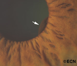
A 64-year-old female was referred to The New York Eye Cancer Center with a small iris tumor visible at the pupillary margin
Impression
Iris Pigment Epithelial Cyst
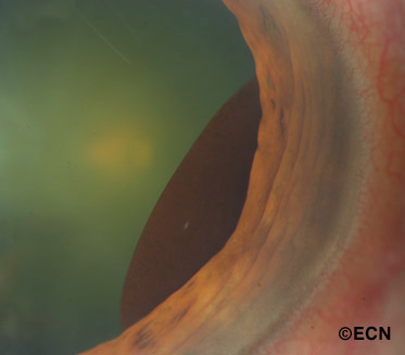
Iris pigment epithelial cysts are commonly located at the iridociliary junction and are round or oval. When observed by slit-lamp examination, they have a smooth brown surface. Visualization of the tumor can be improved by dilation of the pupil.
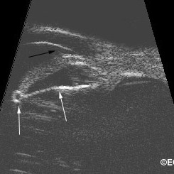
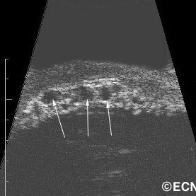
Iris pigment epithelial cysts have thin walls and sonolucent contents (as imaged by UBM). It is generally accepted that the high reflectivity of the cyst wall is caused by its epithelial cell lining and that its sonolucent core is consistent with a liquid content.
Iridociliary cysts typically displace the iris root anteriorly. This can induce a focal plateau-iris configuration with or without angle-closure. Though single cysts are more common, multiple cysts are found in at least one third of cases. When multiple cysts involve more than 180 degrees of the iris, as it does in 10% of patients, angle-closure glaucoma may develop.
The natural history of iris pigment epithelial cysts is poorly understood. Therefore serial observation is warranted.
Recommendation:
- Observation
- Laser cystotomy for progressive angle closure, threatening acute glaucoma
References
- Differential Diagnosis of Anterior Segment Cysts by Ultrasound Biomicroscopy. Authors: Marigo FA, Esaki K, Finger PT, Greenfield DS, Liebmann JM, Ritch R. Ophthalmology 1999;106:2131-35
- Anterior Segment Tumors: Current Concepts and Innovations. Marigo FA, Finger PT. Survey of Ophthalmology 2003;48:569-593.
- Transpupillary Nd:YAG laser cystotomy for iris pigment epithelial cysts with secondary progressive angle closure. Ophthalmic surgery, lasers & imaging [1542-8877] Kathil, Pratima. yr:2011 vol:42 Online pg:e40 -e43


