Finger’s Essential Ophthalmic Oncology Textbook is Now Open-Access for the Public!Dr. Finger Presents the Zimmerman Lecture at the AAO SymposiumECF Supported Research Featured in New York Eye and Ear Infirmary 200 Years CelebrationThe Garg-Finger Staging System for Retina Capillary Hemangioma
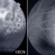
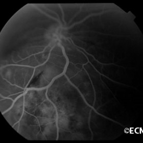
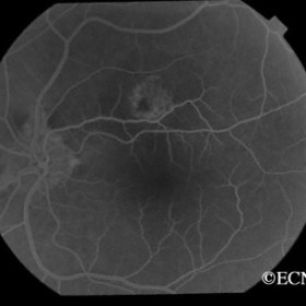
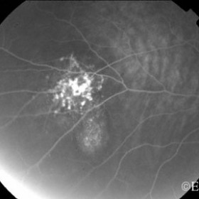
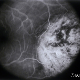
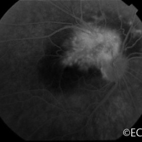
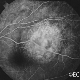
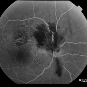
Fluorescein/ICG Angiography Choroidal HemangiomaLeft: Fluorescein angiography reveals the coarse vascular pattern that is commonly found on the tumor surface. Right: This pattern disappears in the late phases and is replaced by nonspecific diffuse hyperfluorescence.Choroidal metastasis - Wilm`s tumorChoroidal metastasis - Wilm`s tumor - Note irregular tumor filling and no definite tumor vessels.Choroidal NevusChoroidal Nevus - Focal fluorescence documented over the apex of this choroidal nevus during angiography.Choroidal NevusChoroidal Nevus- Note the small exudative retinal detachment along the inferior margin of this suspicious choroidal nevus.Circumscribed Choroidal HemangiomaCircumscribed Choroidal Hemangioma - A course vascular pattern is best seen with fluorescein angiography.Combined hamartomaCombined hamartoma of the retinal pigment epitheliumMetastatic choroidal tumorMetastatic choroidal tumor - Note the diffuse fluorescence and lack of well-defined tumor vessels.Radiation optic neuropathyRadiation optic neuropathy - Hemorrhagic variant.