By Paul T. Finger, MD
History
A 63 year old male was referred to The New York Eye Cancer Center with a 6 month history of progressive painless proptosis of the right eye.
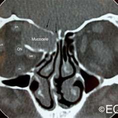
These magnetic resonance imaging (MRI) studies demonstrate displacement of the optic nerve, a bright T1 tumor image, and a variably bright T2 tumor image. The tumor is noted to involve the orbit, the ethmoid and frontal sinuses.
Despite this large orbital tumor with optic nerve displacement, the patient was 20/20 OU, he had no visual field defect, and no signs of optic neuropathy. A complete medical survey was initiated and the patient was cleared for surgery. A combination of anterior orbitotomy and transnasal ethmoidectomy were performed to evacuate the mucus and allow for future drainage.
Though recurrence is possible, this surgery is typically curative. We recommend sending the mucoid contents for culture and sensitivity and cytopathology.
Impression:
Orbital Mucocele


