Case #8: 3D Ultrasound Topography of Choroidal Melanoma

By Paul T. Finger, MD

History

A View From Above The Tumor as it Regresses. Note that both the basal and apical dimensions shrink over time. Instead of 3 standard measurements (length, width and height) hundreds of surface data points are evaluated.

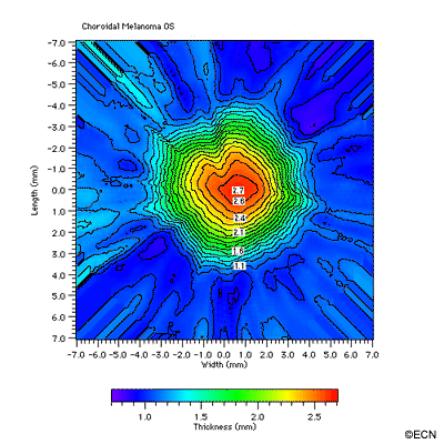
A male with a choroidal melanoma underwent 103Pd (palladium-103) ophthalmic plaque radiation therapy. Three dimensional (3D) ultrasound scans were taken immediately after, 3 and 7 months after treatment. The following renderings were made from that 3D data ultilizing a commercially available 3D ultrasound system.

Clinical Impression

Regressing Choroidal Melanoma

*Note* This patient was treated with palladium-103 ophthalmic plaque radiation therapy.

Comment

An Oblique View of the Tumor as it Regresses. A view from the side of the same tumor. Note that the tumor volume decreases over time. Hundreds of surface data points are evaluated.

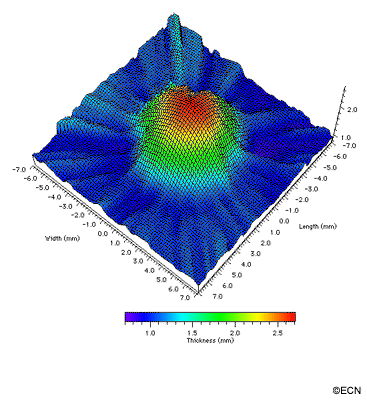
This case clearly demonstrates that choroidal melanomas regress in thickness throughout their volumes. Even though residual pigment may mask regression of the tumors width, 3D topographies clearly demonstrate that these areas of tumor become thinner (as does the tumor’s apex). Topographies should be availabe to evaluate and monitor tumor growth and regression.

Related Links

Patient Stories

"Very well treated by Dr. Finger. He explained everything I needed to know about my issue with detail and attention, putting me at ease and giving me confidence to handle this problem for the rest of my life.”
N.N.

Read More

REQUEST AN APPOINTMENT

Go to Appointment Form

CONNECT AND SHARE