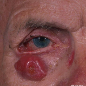
Basal cell carcinoma
Basal cell carcinoma - Right lower eyelid with ectropion and orbital invasion (note the central crater).
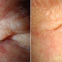
Basal Cell Carcinoma
Basal Cell Carcinoma - A recurrent morpheaform basal cell carcinoma before and six months after external beam radiation therapy.

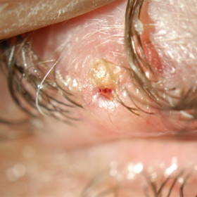
Basal cell carcinoma
Basal cell carcinoma at the medial canthus. Note the pearly margins and central crater.
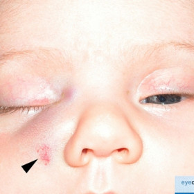
Capillary Hemangioma of Childhood
Capillary Hemangioma of Childhood - Note the epidermal extension (arrowhead), the red cutaneous coloration and mass effect on the nostril.
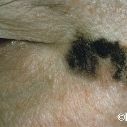
Eyelid Melanoma
Eyelid Melanoma - Pagetoid extension on to the eyelid from malignant melanoma of the conjunctiva.
Hutchinson`s Freckle
Hutchinson`s Freckle - Typically treated as a cutaneous malignant melanoma.
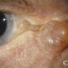
Hydrocystoma at the medial canthus
Hydrocystoma at the medial canthus - Care must be taken to remove all cyst elements and to preserve the lacrimal system.
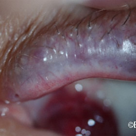
Lympangioma
Lympangioma can involve/invade the eyelids causing bluish discoloration

Lymphangioma of the eyelid
Lymphangioma of the eyelid - A small subcutaneous lymphangiectasia (arrow) of the eyelid extending from a larger orbital lymphangioma.
Malignant melanoma of the eyelid
Malignant melanoma of the eyelid - Note a small diagnostic wedge biopsy was taken prior to excision with large margins.

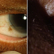
Nevus of Ota
Nevus of Ota - Note the episcleral (ocular melanosis) and eyelid melanosis.
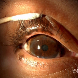
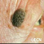
Pigmented nevus
This well circumscribed pigmented nevus does not occlude the punctum and has not grown.
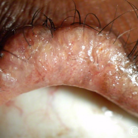
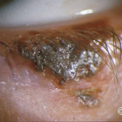
Sebaceous carcinoma (diffuse)
Sebaceous carcinoma (diffuse) in the upper eyelid - Note the eyelash loss and yellow appearance
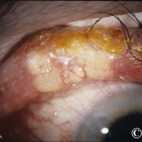
Sebaceous carcinoma in the upper eyelid
Sebaceous carcinoma in the upper eyelid - Note the eyelash loss and chalazion-like appearance.
Seborrheic keratosis
Seborrheic keratosis - not the cobblestone surface, lack of inflammation (dermatitis), and stuck-on appearance.
Squamous Carcinoma of the Eyelid
Squamous Carcinoma of the Eyelid - Note lash loss, erythema, crater and desquamation on its surface.


