By Paul T. Finger, MD
History

A 55-year old female presented to The New York Eye Cancer Center with swelling of her eyelids, signs of scleritis, and pain on eye movement. Her visual acuity was correctable to 20/20 OU, and ophthalmoscopy was significant for a hemorrhagic chorioretinal thickening in the superotemporal quadrant.
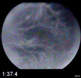
Fundus photographs and a Fluorescein angiogram were obtained utilizing Digital Photography (see right).
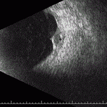
3D-Ultrasonography is significant for a 13 x 13 mm (base), 5.0 mm height choroidal tumor with moderate internal reflectivity. The tumor’s surface is irregularly shaped, the subjacent sclera was thickened, and a retrobulbar lucency “edema” is evident. However, there was no extrascleral tumor extension.
Fluorescein angiography reveals a small patch of intraretinal microangiopathy, leaking retinal vessels, and diffuse areas of blockage.
The differential includes infectious, inflammatory, neovascular, and neoplastic events. When all testing proved negative our options included biopsy or close serial observation.
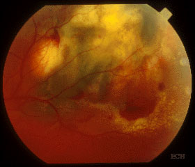
3 Months after Initial Presentation
A fundus photograph (see right) reveals a marked reduction of subretinal fluid and hemorrhage. Exudates have appeared at the infero-temporal margin, and orange pigment on the inferonasal tumor.
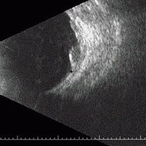
| Serial Ultrasound Evaluations | ||||
| Variable | Presentation | 2 months | 3 months | 5 months |
| Tumor Height | 5.0 mm | 4.0 mm | 1.9 mm | 3.2 mm |
| Reflectivity | Medium | Medium | Variable | Low |
| Surface | Irregular | Irregular | Concave | Dome |
Impression
Necrotic Choroidal Melanoma
This is an atypical presentation for choroidal melanoma. In this case serial observation over time allowed for clearing of the overlying blood and serous fluid. We were eventually able to view of the tumor’s surface (e.g. orange pigment). Serial ultrasonography demonstrated both resolution of the inflammatory and hemorrhagic components, shrinkage, and finally evidence of growth.
In this case, close observation (together with a careful medical work-up) allowed the choroidal melanoma to evolve into a more recognizable form and to demonstrate its tendency to grow.
Recommendation
Ophthalmic Plaque Radiation Therapy
The risks and benefits of continued observation for growth, enucleation, radiation therapy (both proton versus plaque), as well as currently available alternative therapies were discussed in detail. After a discussion of the comparative dosimetric advantages of palladium-103 versus iodine-125 plaque radiation therapy, she chose palladium-103 as treatment for her choroidal melanoma.


