Case #19: Transstromal Iris Pigment Epithelial Cyst
By Paul T. Finger, MD
History
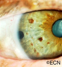
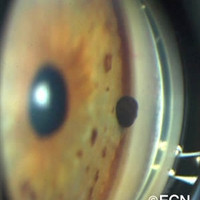
This digital image shows a rounded pigmented tumor extending from the anterior chamber angle.
This patient was noted to have a pigmented tumor on his right iris.
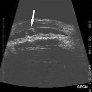
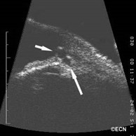
The digital images below will show a rounded pigmented tumor extending from the anterior chamber angle. Gonioscopy revealed no evidence of pigment dusting around the tumor or tumor in the adjacent ciliary body. It was also significant that there was no evidence of distortion of the iris stroma, ectropion uveae, sector cataract or abnormal vascularity (as might be seen with iris melanoma).
"Very well treated by Dr. Finger. He explained everything I needed to know about my issue with detail and attention, putting me at ease and giving me confidence to handle this problem for the rest of my life.”
– N.N.