By Paul T. Finger, MD
History#1
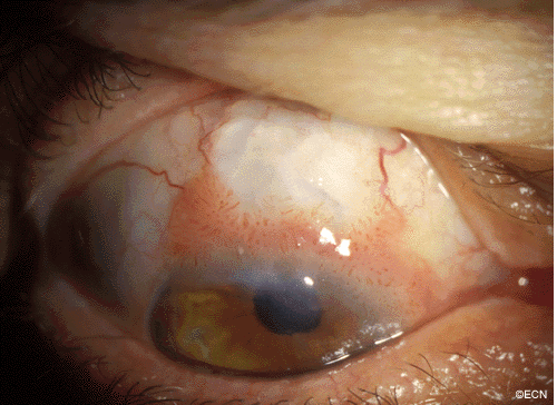 A man with a past ocular history of glaucoma (status-post trabeculectomy) and macular degeneration was referred to The New York Eye Cancer Center with squamous conjunctival neoplasia affecting his right eye.
One might imagine that he is at high risk for intraocular infiltration of his tumor.
Impression
Squamous Conjunctival Neoplasia
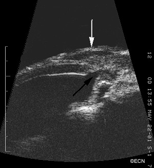
Note: With NO evidence of intraocular invasion, this patient was treated by tumor resection, superficial keratectomy, and adjuvant cryotherapy (to the episclera and conjunctival margins).
Comment: This case presents an unusual case of squamous conjunctival neoplasia that had grown over a functioning filtering bleb.
Findings on high-frequency ultrasound and the presence of iris neovascularization can be helpful in determining if squamous conjunctival neoplasia has infiltrated the eye.


