By Paul T. Finger, MD
History
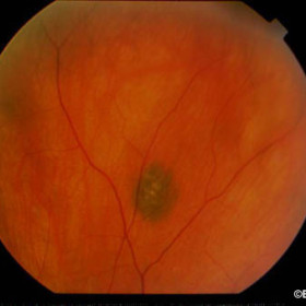
In April of 2001, a 50-year-old female was referred for evaluation of a pigmented tumor in her right eye.
Unlike a choroidal melanoma this choroidal nevus did not have orange pigment, subretinal fluid or thickness greater than 2 mm. There were drusen over the area of pigmentation.
Impression
Choroidal Nevus
Comment
In cases of choroidal nevi, it is important to take a fundus photograph which serves as a baseline for future comparison. In this case the borders were clearly defined and a copy of this photograph was sent to the referring physician (to aid in his continued care). Should he repeat this fundus photograph, it will be important to compare the relative intraocular illumination used during photography. Consider the following series of fundus photographs taken during this patient’s initial visit.
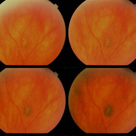
We present this clinical pearl to aid in your continued care of patients with choroidal nevi. Next time you are evaluating a fundus photograph and think there is tumor growth, double check the background illumination to make sure the two pictures were taken at similar settings.


