By Paul T. Finger, MD
History
This patient was also noted to have a pigmented iris tumor in his right eye.
Impression
Posterior Iris Pigment Epithelial Cyst

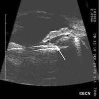
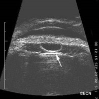
Iris pigment epithelial cysts are typically located behind the iris stroma. As they expand, the cyst wall comes in contact with the natural lens (or IOL) posteriorly and push the iris stroma anteriorly. Gonioscopy and high-frequency ultrasound examination usually reveals focal angle closure. Since most of the trabecular meshwork remains open, most patients do not develop narrow angle glaucoma.
Giant iris pigment epithelial cysts can cause total angle closure warranting intervention. These patients have been treated with Yag-laser iridotomies or surgical iridectomy.
Almost all iris pigment epithelial cysts can be monitored with periodic observation for evidence of secondary glaucoma. Slit-lamp photography, gonioscopy and high-frequency ultrasonography are particularly helpful in evaluating for progressive enlargement. The natural history of iris pigment epithelial cysts is poorly understood.


