By Paul T. Finger, MD
History
An 80-year-old male with a past history of coronary artery disease,and cataracts (removed in 1986 and 1996). He has been taking aspirin and Vitamin E for several months, but had discontinued both due to ecchymosis on both hands.
He was referred to The New York Eye Cancer Center for an evaluation of an intraocular tumor and 2 weeks of ocular pain with photophobia OD.
Ophthalmic examination revealed a visual acuity of 20/25 OU. His pupils were asymmetric (without APD) and his visual fields were restricted OD, full OS. Ocular motility was within normal limits.
Intraocular pressure measurements were 13 OD and 11 OS. Slit lamp biomicroscopy revealed posterior chamber implants with some retained cortical material in the superotemporal quadrant OD. There is no tumor in the angle on gonioscopy and no mass effect on the iris. Both high frequency and low frequency ultrasonography were performed:
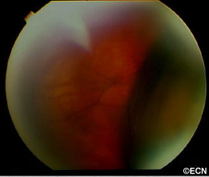
A 2-dimensional 10 MHz transocular ultrasound at the plane of the equator-anterior meridian revealed a multilobular thickening of the anterior choroid with variably reflective subretinal echoes. On dynamic ultrasound examination of the retina, it was found to undulate with eye movement (consistent with hemorrhagic choroidals). A-scan was used to evaluate the subretinal material. It was found to be completely echolucent (ARROW) at 67 db consistent with blood or (less-likely) exudative fluid.
Impression
Spontaneous Hemorrhagic Choroidals
Recommendations
Dr. Finger’s primary recommendation was serial observation. There was a small possibility of a hemorrhagic neoplasm beneath the choroidals or a ring melanoma. With this in mind, the patient was given a complete metastatic survey as well as evaluations for coagulopathy. He was advised not to lift, strain, or rub his eye, that he should use Tylenol instead of aspirin, and not use his Vitamin E supplements. He was warned that should the blood dissect into his macula, this may result in some level of permanent (unilateral) loss of vision. He was to continue taking Advil for pain control. One month later, along with his pain the choroidals were largely resolved. His vision continued to be 20/25 OU.


