Day 5: Live From the ISOO 2019!

Day 5 at the ISOO: Corneal & Conjunctival Cancers

Every two years, eye cancer specialists worldwide convene at the International Society of Ocular Oncology Conference in order to review latest research and encourage international collaboration within the field of ophthalmic oncology.

For details on the ISOO Biennial Conference, read more here.


The final day of the ISOO was dedicated solely to cancers of the conjunctiva, lid, and orbit. Another day filled with research findings and discussion on proper treatment and lesser-known, evidently effective therapeutic techniques.


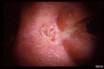
OSSN: Is Surgery Really Necessary?

Poster by Dr. Paul T. Finger and Dr. Chaugule

Is surgery necessary? Dr. Sonal Chaugule asked the question in the paper, Topical chemotherapy for giant ocular surface squamous neoplasia of the conjunctiva and cornea, published alongside Dr. Paul Finger and Dr. Jennifer Park. In the case of ocular surface squamous neoplasia (OSSN), the standard of treatment for years has been surgical excision with adjuvant cryotherapy. However, Chaugule and Finger performed this study in order to compare chemotherapeutic eye drops versus the surgical standard. The eye drops alone have the benefits of convenience and avoidance of complications (however unlikely) from surgery. Ultimately, chemotherapy was found to be sufficient treatment for even giant OSSN. The found that “ topical therapy treated the entire ocular surface and avoided surgical intervention … topical chemotherapy [also] resulted in less scarring and stem cell loss compared to [surgery]. Clearly, larger tumors than widely expected can be controlled with topical chemotherapy alone.”

The full text is available, public-access here.


Palladium Succeeds with Monocular Patient

From Left: Dr. Ekaterina Semenova, Dr. Paul Finger

Our now-graduated fellow Dr. Ekaterina Semenova presented on an already-blogged about, very unique case. A patient had only one functioning eye, and had previously undergone a corneal transplant in this eye. After treating this patient with Palladium-103 plaque brachytherapy through the natural and transplanted cornea, the tumor was found to be controlled and the patient’s vision was conserved.

Read about this case in greater detail on our blog here.


Super-Thick Amniotic Membrane Grafts for Eye Cancer Patients

From Left: Dr. Paul Finger, Dr. Puneet Jain

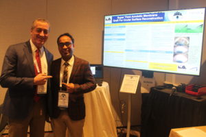
Another ECF graduate, Dr. Puneet Jain, presented on Super Thick Amniotic Membrane Graft (ST-AMG) Successfully Used for Ocular Surface Reconstruction. Compared to the often-used single-layer amniotic membrane graft, the larger grafts allow for greater patient comfort, as well as greater ease of suture, increased likelihood of remaining in place for the required duration, as well as decreased likelihood of tearing. 

Read about this case in greater detail on our blog here.


Keratoacanthoma: An Unusual Location!

A fellow of Dr. Santosh Honavar at The Centre for Sight in Hyderabad, India, Dr. Ankit Singh Tomar will be joining us as the most recent Eye Cancer Foundation fellow. He presented a unique case of a conjunctival keratoacanthoma on the tarsal conjunctiva. It was the first reported case of this extremely unique pathology, and was managed surgically without complications at The Centre for Sight.

We hope to see more great research from Dr. Tomar after he joins us for his fellowship training in ophthalmic oncology her at The New York Eye Cancer Center.

Welcome to our new fellow, Dr. Tomar!


The End, For Now

This marks the final day of the conference and it has since come to a close, though many plans have been made for international cooperation, multi-center databases, and collaborative research so that eye cancer patients worldwide will one day not only have access to care, but have access to the best care we have available. Progress is being made behind the scenes as we speak, and we hope to bring you amazing news for the next conference in 2021.

Until then, thank you for your attention and we thank the amazing physicians and researchers for their great work.

Dr. Finger with delegates from Russia
Dr. Finger with our most recent ECF fellowship graduate, Dr. Ann Musika (right) and her mentor, Dr. Vicktoria Dai (left)

Remember — you can always be a part of our research and international fellowship training by giving a gift to The Eye Cancer Foundation. Dozens of papers this year were presented due to funding from The ECF.


Donate Now!


Malignant Melanoma of the Eyelid

By Paul T. Finger, MD

Description

Melanoma of the eyelid is a relatively rare tumor making up less than 1% of eyelid cancers. However, like other skin melanomas it can spread to other parts of the body. This tumor typically appears as a pigmented thickening (tumor) of the eyelid or extension of pigment from the conjunctiva. It is characterized by a patient history of or photographically documented growth.

Symptoms

Malignant melanoma of the eyelid may look like an eyelid nevus, but it more likely to be  variably pigmented, change color, bleed and/or grow. All pigmented eyelid tumors of the eyelid should be evaluated by an eye cancer specialist.

Diagnosis

All eyelid tumors should be photographed for comparison with future examinations. if possible, patients should keep a copy of their baseline photographs. Malignant melanoma of the eyelid is distinguished from an eyelid nevus in that it can be variably pigmented, change color, bleed and/or grow. If your doctor makes a clinical diagnosis of eyelid melanoma or change is noted, the tumor should be evaluated by an incisional (not superficial) biopsy. This is because the depth of the tumor is an important risk factor for spread to the rest of the body.  A simple wedge biopsy can be sent for pathologic evaluation to aid in the diagnosis. To evaluate the entire tumor, primary excisional biopsies need be performed. Once the diagnosis is made on pathology, systemic staging with total body, scalp to toes PET/CT imaging should be performed.

Treatments

When the pathology diagnosis confirms or discovers that the tumor is malignant melanoma of the eyelid (eyelid cancer), a medical work-up is ordered to stage the patient and thus rule out metastatic spread to other parts of the body. If there is no metastatic disease, the tumor can be surgically removed (including large margins of normal appearing tissue). At the time of surgery, some doctors will also remove regional lymph nodes near the tumor (in order to determine if the tumor has locally spread).

Ophthalmic plastic surgery techniques are usually required to repair the defect caused by tumor removal. The extent of surgery is governed by the ocular distribution of the tumor. That said, it can range from simple eyelid resection (wedge or Moh’s) to removal of the eyelid, debulking the orbit, to orbital exenteration. Radiation (external beam or brachytherapy implant) may be added if surgical margins are not possible.  Most all patients can be repaired using one of these techniques (see Treatment Types Section).

Related links


Basal Cell Carcinoma (Eyelid Cancer)

By Paul T. Finger, MD

Description

A basal cell carcinoma anterior to the medial canthus: Note the pearly margins and the central crater.

The most common type of eyelid cancer is basal cell carcinoma. Most basal cell carcinomas can be removed with surgery. However, many older patients will try to ignore these slow growing tumors. However, it is important to know that when left  untreated, these tumors can grow around the eye and into the orbit, sinuses and brain. Basal cell carcinomas are more commonly found on the lower eyelids and almost never spread to other parts of the body (metastasize).

Symptoms

Patients with basal cell carcinomas most commonly notice a reddish nodule slowly forming on their eyelid. The tumor is most commonly found on the lower eyelid, followed by the medial canthus (skin toward the nose) and can occur on the upper eyelid. Eyelash loss (around the tumor) suggests that a basal cell carcinoma is malignant.

Less commonly, basal cell cancers can be pigmented or present without any nodule at all. When the tumor does not make a nodule and grows within the eyelid, it can induce pulling of the eyelid (away from the eye). Doctors call this form of basal cell carcinoma “morpheaform,” which are much more difficult to cure because its edges are harder to define.

Diagnosis

Though small tumors can be photographed and followed for evidence of growth (prior to biopsy); once your eye cancer specialist suspects basal cell carcinoma, most eye cancer specialists will suggest tumor biopsy. This specimen is sent to the pathologist to confirm the diagnosis prior to complete removal or treatment of the tumor. Biopsies can be performed in the doctor’s office, or in the operating room prior to definitive treatment.

Treatments

Once the diagnosis is confirmed by the pathologist, treatment will be recommended. Wedge resection and Moh’s surgery require the surgeon continue to remove the tumor until the margins (edges) are negative (free of tumor). Unlike most skin, the eyelid are a complex functional apparatus that requires special reconstruction techniques.

Most basal cell carcinomas are cured when they are small. Unfortunately, some patients choose to ignore or deny the existence of these tumors. Those patients allow their tumor to invade behind the eye and become difficult or impossible to remove. In these cases, combinations of surgery, radiation and chemotherapy may be required to control or destroy the tumor.

Additional Info

This is a sun related cancer. Dr. Finger suggests, “Think of sunglasses as sunblock for your eyes”™

Related Links

Search Recent Scientific Publications about Basal Cell Carcinoma of the Eyelids


Patient Stories

"Very well treated by Dr. Finger. He explained everything I needed to know about my issue with detail and attention, putting me at ease and giving me confidence to handle this problem for the rest of my life.”
N.N.

Read More

REQUEST AN APPOINTMENT

Go to Appointment Form

CONNECT AND SHARE