Ocular Melanoma Mystery: Rare Eye Cancer Found in 36 Auburn University Graduates


With 87,110 diagnoses estimated to be made in 2018 for U.S. Americans, skin cancer, particularly a melanoma, is the most frequently diagnosed cancer in the United States. Ocular melanomas, however, remain uncommonly diagnosed, affecting just six in every one million people a year. Given the extreme rarity of ocular melanomas, doctors and researchers were shocked to find this disease found in highly concentrated numbers in two states. A total of 36 people — all graduates from Auburn University, Alabama — were diagnosed with ocular melanoma. From Huntersville, North Carolina, 18 patients were also found out to have the same disease.

Left to right: Ashley McCrary, Allison Allred, Juleigh Green and Lori Lee

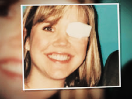
Juleigh Green, Allison Allred, and Ashley McCrary had spent their years at Auburn University as close friends. And later on in their lives, each woman discovered they had the same rare disease. Juleigh Green was the first among her friends to be diagnosed. Just 27 at the time, Green was experiencing strange flashes of light obstructing her vision and consulted her ophthalmologist immediately. In an interview with CBS, Green explains her shock upon what they found:

“[My doctor] said, ‘There’s a mass there, there’s something there, I don’t know what it is, but it looks like it could be, you know, a tumor,’” Green said. “It’s like you had the breath knocked out of you, you know?”

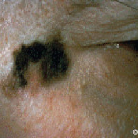
Allison Allred, who also was experiencing flashes of light for 7 to 10 days, was the second of their friend circle to be diagnosed in 2001, at the age of 31. Her doctor had first believed that the flashes were due to a retinal detachment. According to Allred, her doctor had said: “Well, [the retina] is detached because there’s a 10 millitmeter melanoma sitting on it.”

Both Green and Allred opted for enucleation and have had their afflicted eye removed. However, Allred’s extremely aggressive and stubborn cancer has since recurred nine times in six separate places in her body. “Two days ago I found out that it’s come back to my brain,” Allred told CBS, “So, I’m actually gonna have radiation on my brain tomorrow.”

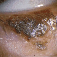
Ashley McCracy was the third friend, with her own diagnosis coming from observing unusual black spots along her iris. In an interview with CBS News correspondent Anna Werner, McCrary said:

“What’s crazy is literally standing there, I was like, ‘Well, I know two people who’ve had this cancer.”

“And did you understand then how strange that was?” asked Werner.

“No. No, I didn’t.”

All three women were treated at Sidney Kimmel Cancer Center at Thomas Jefferson University in Philadelphia, Pennsylvania. McCrary mentioned Green and Allred’s similar diagnosis to her oncologist at Kimmel Center, Dr. Marlana Orloff.

Orloff was baffled.

“Most people don’t know anyone with this disease,” Orloff said. “We said, ‘OK, these girls were in this location, they were all definitively diagnosed with this very rare cancer — what’s going on?”

A fourth Auburn alumna, Lori Lee, is also being treated at Kimmel Center. “This is a rare cancer, so it’s not like you can just go anywhere and have anybody know anything really about it,” Lee said. “Until we get more research into this, then we’re not gonna get anywhere. We’ve got to have it so that we can start linking all of them together to try and find a cause, and then one day, hopefully, a cure.”

Orloff and her fellow researchers and oncologists at Kimmel Center immediately began to investigate this bizarre case. Thus far, the Alabama Department of Health states that “it would be premature to determine that a cancer cluster exists in the area”. Officials at Auburn University hope that research will help illuminate the cause of this rare cancer appearing at such high concentrations in Alabama and North Carolina.

Each patient’s emotional response to this mystery cannot be understated. “That was very hard for me,” McCrary told CBS. “Growing up, the one thing that I liked about myself was my eyes.”

Juleigh Green, pictured above, was the first of her two friends from Auburn University to be diagnosed with ocular melanoma.

McCrary’s personal journey in dealing with her cancer diagnosis lead to the creation of the Auburn University Ocular Melanoma Page on Facebook, which has astoundingly discovered 36 more graduates afflicted with ocular melanoma. The Facebook Page offers itself as an effective support network for these graduates.

Kimmel Center researchers continue to look for answers to explain these phenomena both in Alabama and North Carolina.

Stay tuned for the latest updates on this case and others by keeping eyecancer.com in your bookmarks.


Patient Stories: Maureen’s Journey

As part of an ongoing initiative from the Eye Cancer Foundation, The ECF accepts submissions where patients share their personal story through diagnosis and treatment. Of course, the ECF is always accepting submissions for Patient Stories and would love to hear from you. Below is the first publication, sent in by a patient by the name of Maureen. 

In December 1994, I went to see my optometrist, Dr. Quist, at Pildes in New York. I was going to Boston for New Year’s and my contacts were bothering me. Well, Dr. Quist pointed out that I had not had a good exam in a while and she wanted to do one. I said sure.

As she studied my left eye, she took a bit more time than she did with the right eye. Afterward, she wrote out a note, put it in a sealed envelope, and said that I should see my ophthalmologist. That was the beginning.

Upon consulting my ophthalmologist, Dr. Muldoon, I discovered that the little freckle Dr. Quist luckily saw was a possible cancerous condition which would have to be monitored. In May 1995, I went for what I thought would be another routine visit with Dr. Muldoon. It was not routine. After the exam, he asked his receptionist to see if Dr. Finger was in the hospital that day. He was, and I was sent down to his office at the New York Eye and Ear Infirmary. It was only when I saw the words Melanoma Study beside the door that I actually realized: Ah. Not just cancer, but melanoma.

In the blink of an eye, so to speak, life changed. I was immediately sent for the first of ever-so-many fluorescein injections and pictures, ultrasounds — the works. Dr. Finger observed the nevus until November 1995 and then decided that treatment was needed. At the time of all this I was a volunteer at Sloan-Kettering and I knew how helpful it was to speak to patient-to-patient volunteers. Well, there weren’t any; the ailment was so rare. My medical doctor, who had practiced for years, had never heard of it and had to research it. While having experience seeing the courage of the cancer patients I worked with, and the research being done with all sorts of cancers, I myself was scared enough with my disease to determine that I would go with enucleation for treatment. The idea of the melanoma was very upsetting to me.

Dr. Finger set me up with an appointment at Johns Hopkins’ Wilmer Institute, where they were doing the national study on ocular melanoma. My husband and I went to Baltimore for a consultation and I was grateful that I took the opportunity to participate in research. The doctor did the exam, discussed his findings and – what I remember most clearly – discussed prosthesis. The reality of removing the eye (which he was, in our opinion, recommending) was what I heard, and it was the dose of reality I needed.

In New York, my husband accompanied me to my next consultation with Dr. Finger. I had not involved him directly up until then – perhaps it was a way of not really acknowledging what was happening. But, Dr. Finger then launched into a description of Palladium-103, the isotope he planned to use to radiate the tumor, with hopefully a good chance at not affecting the surrounding tissue. My tumor was a choroidal melanoma.

I went for it. I was in the New York Eye and Ear Infirmary for 8 days – one day longer than estimated. While I was disappointed to have to stay even one day longer, in retrospect, the nursing care was much better than handling treatment on my own. From my room I had a view of the Empire State lit up for Christmas, and I still see that building out of my office window every day — with both eyes. I was released on December 13, 1995 – St. Lucy’s Day – Santa Lucia – the day celebrated as the festival of light.

I am fortunate to have had a good recovery and to have retained my eyesight. As I say, so far so good, and still, I go to each 6 month follow up with caution. I don’t think anyone would be arrogant enough to forget the trauma. I am grateful to this day to the two patients whom Dr. Finger put me in touch with, who spoke to me before treatment and shared their experiences. Only someone who has been there truly knows what the experience feels like. I have made two wonderful friends who took the road after I did.

Meeting with the fine, young doctors who are being trained through The Eye Cancer Foundation especially is truly inspiring. And I know that patients today have a wide support network with people who share so generously and bravely.


Malignant Melanoma of the Eyelid

By Paul T. Finger, MD

Description

Melanoma of the eyelid is a relatively rare tumor making up less than 1% of eyelid cancers. However, like other skin melanomas it can spread to other parts of the body. This tumor typically appears as a pigmented thickening (tumor) of the eyelid or extension of pigment from the conjunctiva. It is characterized by a patient history of or photographically documented growth.

Symptoms

Malignant melanoma of the eyelid may look like an eyelid nevus, but it more likely to be  variably pigmented, change color, bleed and/or grow. All pigmented eyelid tumors of the eyelid should be evaluated by an eye cancer specialist.

Diagnosis

All eyelid tumors should be photographed for comparison with future examinations. if possible, patients should keep a copy of their baseline photographs. Malignant melanoma of the eyelid is distinguished from an eyelid nevus in that it can be variably pigmented, change color, bleed and/or grow. If your doctor makes a clinical diagnosis of eyelid melanoma or change is noted, the tumor should be evaluated by an incisional (not superficial) biopsy. This is because the depth of the tumor is an important risk factor for spread to the rest of the body.  A simple wedge biopsy can be sent for pathologic evaluation to aid in the diagnosis. To evaluate the entire tumor, primary excisional biopsies need be performed. Once the diagnosis is made on pathology, systemic staging with total body, scalp to toes PET/CT imaging should be performed.

Treatments

When the pathology diagnosis confirms or discovers that the tumor is malignant melanoma of the eyelid (eyelid cancer), a medical work-up is ordered to stage the patient and thus rule out metastatic spread to other parts of the body. If there is no metastatic disease, the tumor can be surgically removed (including large margins of normal appearing tissue). At the time of surgery, some doctors will also remove regional lymph nodes near the tumor (in order to determine if the tumor has locally spread).

Ophthalmic plastic surgery techniques are usually required to repair the defect caused by tumor removal. The extent of surgery is governed by the ocular distribution of the tumor. That said, it can range from simple eyelid resection (wedge or Moh’s) to removal of the eyelid, debulking the orbit, to orbital exenteration. Radiation (external beam or brachytherapy implant) may be added if surgical margins are not possible.  Most all patients can be repaired using one of these techniques (see Treatment Types Section).

Related links


Patient Stories

"Very well treated by Dr. Finger. He explained everything I needed to know about my issue with detail and attention, putting me at ease and giving me confidence to handle this problem for the rest of my life.”
N.N.

Read More

REQUEST AN APPOINTMENT

Go to Appointment Form

CONNECT AND SHARE