Adenoid Cystic Carcinoma of the Lacrimal Gland

By Paul T. Finger, MD

Description

Computed Radiographic Tomography (CT) demonstrates and Adenoid Cystic Carcinoma of the Lacrimal Gland with Orbital Extension (arrow)
Adenoid cystic carcinoma is a type of cancer that affects glandular structures. Around the eye there is a lacrimal (lac-kree-mall) gland that makes tears. Orbital adenoid cystic carcinoma usually occurs in patients 20-50 years old.

Symptoms

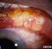
When an adenoid cystic carcinoma of the lacrimal gland grows, it typically pushes the eye down, towards the nose and forward. It can cause bulging of the eye (called proptosis). Another characteristic of adenoid cystic carcinoma is that it also invades local nerves causing pain. Therefore, pain and bulging of the eye are the most common symptoms of adenoid cystic carcinoma of the lacrimal gland.

Diagnosis

A complete eye examination with a clinical history and ophthalmic examination are crucial to the diagnosis of adenoid cystic carcinoma of the lacrimal gland and orbit.

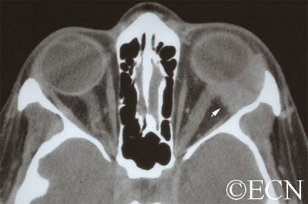
CAT scans, MRI’s and ultrasounds are also helpful in determining the diagnosis. When the eye cancer specialist sees a well-defined tumor in the superior-temporal (upper – outer) part of the orbit, that may have eroded into adjacent bone and/or extend into the orbital apex, he or she should suspect the tumor might be an adenoid cystic carcinoma.

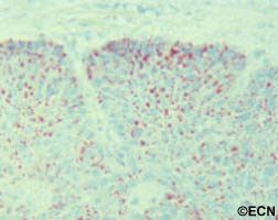
Other tumors to consider include: benign mixed tumor, adenocarcinoma, dacryoadenitis, or other processes affecting the lacrimal gland. Often times a biopsy is need to confirm the clinical diagnosis.

Treatments

When possible, an adenoid cystic carcinoma should be totally removed. This usually requires a surgery called a lateral orbitotomy. Unfortunately, total removal is often impossible due to the tumor’s size, shape, and presence of invasion. Should the adenoid cystic carcinoma be found to have a capsule, and should your doctor be able to remove it within its “capsule,” total excisional surgery offers the best prognosis.

However, in many cases the adenoid cystic carcinoma extends beyond the capsule making removal of the entire adenoid cystic carcinoma nearly impossible. It may require removal of the orbital contents, bones and adjacent structures. Due to the poor (local control) results from this type of extensive surgery; combinations of surgical removal, regional chemotherapy and local radiation therapy may be offered as an alternative.

Additional info


Sebaceous Carcinoma of the Eyelid

By Paul T. Finger, MD

Description

Sebaceous carcinoma arises from the glands within the eyelids, caruncle or eyebrow. They are more commonly found on the upper eyelid and in middle-aged patients.

Symptoms

Sebaceous cell carcinoma is suspected due to evidence of eyelash loss and the formation of a yellow-nodule. This tumor can also present as a persistent (months) non-responsive blepharitis or conjunctivitis. In these cases, a high index of suspicion for sebaceous cell carcinoma will lead to biopsy and the diagnosis. Once sebaceous carcinoma is suspected a biopsy is warranted. Before surgery, the pathologist should be advised of this possible diagnosis so the specimen can be processed appropriately

Sebaceous carcinomas are one of the rarest eye cancers and can look like a chalazion (stye) or conjunctivits. In that sebaceous carcinoma can mimick these relatively benign dieases, eye care specialists should be suspicious of this tumor in any patient with persistent conjunctivitis, blepharoconjunctivitis or chronic/recurrent chalazion. Therefore, any conjunctivitis or chalazion that is not getting better after 3 months of observation, should be biopsied.

Diagnosis

Sebaceous carcinoma of the eyelid typically presents as a yellow-nodule in the upper lid. The patient may have history of colon, breast, genitourinary or other skin malignancies (Muir-Torre Syndrome).* When a nodular tumor, it can cause eyelash loss, but is not painful (compared to styes). This tumor can also present as a hypervascular conjunctival tumor and tends to spread along the conjunctival surface. In these cases sebaceous carcinoma looks like conjunctivitis.

It is worth repeating that “any conjunctivitis or chalazion that is not getting better after 3 months of observation should be biopsied.” If cytopathology is available at your medical center, a scrape biopsy of the conjunctiva can be initially performed in the office setting. However, the gold standard for diagnosis of sebaceous carcinoma is histopathologic evaluation of a fresh tissue specimen.

Once the diagnosis is made, a metastatic survey is warranted. Sebaceous carcinoma can spread to regional lymph nodes (pre-auricular and cervical) as well as to lungs, brain, liver and bone. At The New York Eye Cancer Center, we obtain a total body PET/CT scan (from head to toes) for staging. Published series have reported that the occurrence of metastatic disease was dependent on the size and location of the primary tumor and occurred in up to 27% of patients. Local tumor invasion of the lymph nodes, orbit or metastatic sites were associated with a poor prognosis for survival.

Treatments

Biopsy usually requires a full thickness lid section sent for histopathologic evaluation prior to formalin fixation. Histopathologic evaluation reveals foamy cytoplasm (with hematoxylin and eosin) This specimen is found to be oil-red-o positive, consistent with sebaceous carcinoma. *note* Sebaceous carcinomas may occur several years before a gastrointestinal malignancy. This occurrence has been described as the autosomal dominant - Muir-Torre Syndrome.
Biopsy usually requires a full thickness lid section sent for histopathologic evaluation prior to formalin fixation. Histopathologic evaluation reveals foamy cytoplasm (with hematoxylin and eosin) This specimen is found to be oil-red-o positive, consistent with sebaceous carcinoma. *note* Sebaceous carcinomas may occur several years before a gastrointestinal malignancy. This occurrence has been described as the autosomal dominant – Muir-Torre Syndrome.

Treatment requires a resection of the malignancy. Frozen section control can be used to provide negative margins. Larger surgeries, cryodestruction and radiation may be required if resection is not possible. Exenteration (complete removal of the orbital contents is sometimes required for extensive or recurrent disease).

* Muir-Torre Syndrome: A rare, genetically autosomal dominant cancer syndrome. Patients with Muir-Torre can develop cancer of the colon, breast, genitourinary tract, skin and eye lesions, including keratoacanthomas and sebaceous carcinoma.

Related links


Patient Stories

"Very well treated by Dr. Finger. He explained everything I needed to know about my issue with detail and attention, putting me at ease and giving me confidence to handle this problem for the rest of my life.”
N.N.

Read More

REQUEST AN APPOINTMENT

Go to Appointment Form

CONNECT AND SHARE