Optic Nerve Sheath Meningioma

By Paul T. Finger, MD

Graphic of how an eye can look with a posterior orbital tumor.

Orbital and optic nerve meningioma can extend from the brain into the orbit (behind the eye) and push the eye forward causing a bulging of the eye called proptosis. Though rare, when they occur, they are a significant cause of vision loss.

Symptoms

Patient with orbital meningioma typically have proptosis (bulging eye). Optic nerve compression can cause optociliary shunt vessels to form, as well as loss of vision. Depending on the location, size and degree of optic nerve involvement; patients can develop monocular and/or junctional defect is the patients field of vision.

Diagnosis

Opto-ciliary shunt vessels resulting from optic nerve compression

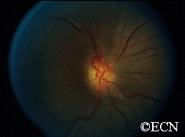
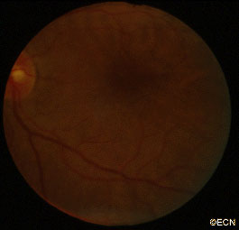
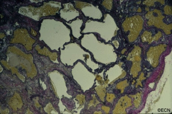
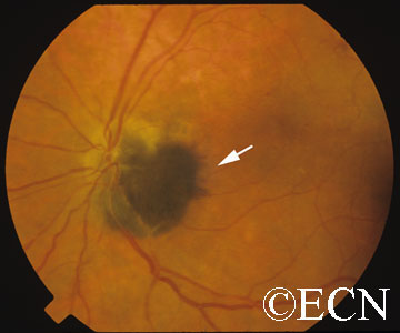
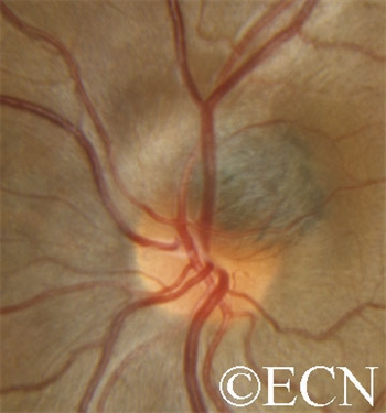
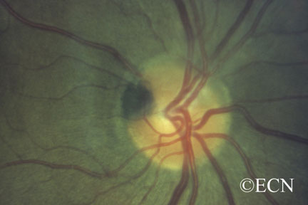
Patients usually present in their 40s and may have neurofibromatosis type 2. In making this diagnosis, one should look for the triad of vision loss, optic atrophy and abnormal vessels on the optic nerve. The nerve head can appear raised. Enlarged blood vessels are called “optociliary shunt vessels” and indicated that the meningioma has disrupted the natural circulation through the optic nerve to the retina and choroid. Angiography of the optic nerve head will clearly demonstrate the abnormal blood vessels. Magnetic resonance imaging (MRI), ultrasonography and computed tomographic (CT) imaging have been used to evaluate the orbital tumor and measure the optic nerve sheath diameter. CT is particularly helpful for imaging calcium within the tumor.


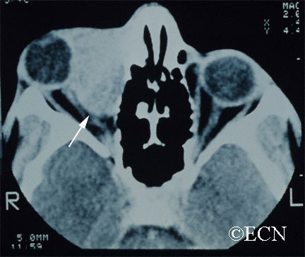
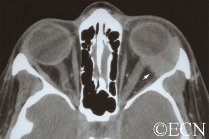
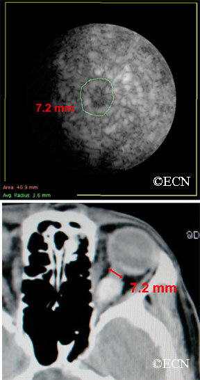
Computed tomography (CT) shows that the eye is pushed forward by this optic nerve sheath meningioma. Notice that the tumor is relatively bright (radio-dense). Imaging of the brain can determine if the meningioma extends into the brain.

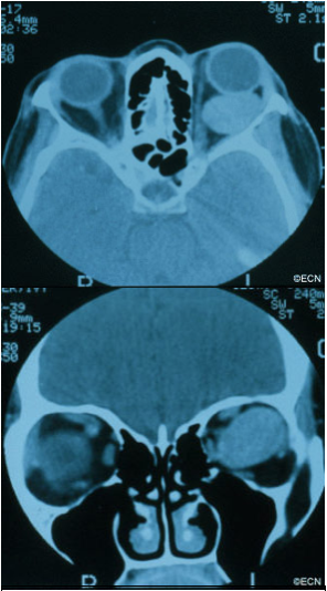
In the images above, note that computed tomography of this optic nerve sheath meningioma. C demonstrates the calcific density of the left optic nerve. An x-ray film shows the linear density (seen on the right side of the film crossing the inferior orbital rim) which corresponds to the optic nerve sheath meningioma seen above.

Coronal "C-scan" ultrasound can be used to compare optic nerve diameters in a case of optic nerve sheath meningioma
Coronal “C-scan” ultrasound can be used to compare optic nerve diameters in a case of optic nerve sheath meningioma

Treatment Plan

Orbital meningioma is typically a slow-growing tumor. Once diagnosed, meningioma can be observed for growth prior to considering intervention. Treatment is indicated when there is a risk of spread to the central nervous system (in primary optic nerve sheath meningioma), documented progressive vision loss, or for rapid growth.

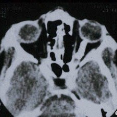
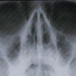
Computed tomography of the optic nerve sheath meningioma. Note the almost calcific density of the left optic nerve. An x-ray film shows the linear density (seen on the right side of the film crossing the inferior orbital rim) which corresponds to the optic nerve sheath meningioma seen above.

Though microsurgical resections have been tried (in an effort to spare the optic nerve), most eventually fail. The goal of local resection should be complete removal of the meningioma. This usually involves removal of the involved optic nerve. If complete surgical removal is not possible or in special circumstances, radiation therapy is commonly employed.

Biopsy:

Indications for biopsy include: atypical tumors, aggressive disease, acute vision loss and when a pathology diagnosis is requested. Certain inflammatory tumors can have a similar appearance to orbital meningiomas. However, biopsy carries risk for vision loss.

Treatment:

Orbital meningioma is typically a slow-growing tumor. One must consider patient age, rate of tumor growth and risk for loss of vision. That said, once diagnosed slow or non-growing meningiomas can be observed for further growth or stabilization prior to considering intervention. In general, treatment is indicated when there is a risk of spread to the central nervous system (in primary optic nerve sheath meningioma), documented progressive vision loss, or for rapid growth.

Treatment alternatives are tailored to the clinical situation. For example, if vision is lost, the tumor is growing toward the central nervous system and is resectable; it is removed. If complete surgical removal is not possible or in special circumstances,  microsurgical resection or external beam radiation therapy can be considered. These decisions are complex and best made with your neuro-ophthalmologist, neurologist and orbital surgeon.

Related Links


Lymphangioma of the Orbit

By Paul T. Finger, MD
Computed Tomography (CT) shows a large lymphangioma (arrow), pushing the eye out of the orbit.

Description

Lymphangioma is rare (less than 7% of childhood orbital tumors). Patients can present with acute proptosis (bulging eye) after minor head trauma, as a gradual proptosis, or after an upper respiratory infection.

Symptoms

Lymphangioma tends to start in the superior and nasal orbital quadrants. More than 50% affect anterior (conjunctival and adnexal) structures. Typically, the lymphangioma bleeds into itself causing cysts of blood (called chocolate-cysts) within the tumor. If the cyst forms behind the eye, it pushes the eye forward. If the tumor forms in the eyelid or structures around the eye “adnexa”, blood filled lymphatic channels called “lymphangiectasias” can be seen beneath the conjunctiva

Diagnosis

A lymphangiectasia is seen beneath the conjunctival surface.

Lymphangioma is usually diagnosed by an eye cancer specialist. A careful history may reveal sudden painful proptosis (bulging of the eye), facial trauma or that the tumor or proptosis started right after a upper respiratory infection.

Physical examination may reveal bluish discoloration of or blood vessels within the eyelid skin. Should the vessels extend under the conjunctiva, they are called lymphangiectasias.

Severe cases can be associated with corneal exposure, ulceration and optic nerve damage.

Treatments

Though lymphangioma patients can present with a history of sudden proptosis (due to bleeding within the tumor), orbital lymphangiomas are typically slow growing. Therefore, most lymphangiomas are followed by observation for growth-related damage as documented by (clinical and radiographic studies) prior to intervention.

Thus, treatment of lymphangioma is indicated when associated with growth, optic nerve compression, corneal exposure problems (keratitis sicca), glaucoma or vision loss.

When treatment of lymphangioma is considered, the goal is rarely complete removal. This is because the edges of most orbital lymphangiomas are poorly defined. When necessary, patients undergo debulking surgeries with a goal of relieving acute optic nerve compression or corneal exposure. In rare cases, orbital lymphangioma patients may require orbital radiation therapy or exenteration for relief of pain.

Related Links


Cavernous Hemangioma of the Orbit

By Paul T. Finger, MD

Description

Computed tomography (CT) – Coronal Section demonstrates displacement of the optic nerve (medially) by the orbital hemangioma.

Hemangioma is a benign tumor that is found to grow within the orbit. Most commonly located behind the eye globe, it can push the eye forward causing eye-bulging doctors call proptosis.

Symptoms

Cavernous hemangioma of the orbit is most commonly seen in middle-aged women. Most are found within the muscle cone, but can be found anywhere in the orbit.

These orbital tumors can indent the back of the eye causing choroidal folds, or push on the optic nerve causing damage (atrophy).

Rarely, the tumor can push the eye so far that the cornea cannot be covered by the eye lids. In these cases, corneal exposure problems (keratitis, superficial punctate keratopathy, ulceration, even perforation) can occur.

Diagnosis

Cavernous hemangioma of the orbit is usually a slow-growing tumor. If the tumor has not damaged the eye, cavernous hemangioma can be observed for growth prior to considering intervention. Should tumor growth occur, it will be measured by eye examinations including (but not limited to) visual acuity, color vision assessment, Hertel exophthalmometry (a measure for proptosis), as well as an evaluation for double vision (strabismus), corneal exposure, retinal damage, vascular damage, and optic neuropathy.

Treatment

Treatment of orbital hemangioma is indicated when there is evidence of growth, optic nerve compression, and corneal exposure (with secondary keratitis sicca), or evidence of vision loss.

The goal of orbitotomy for choroidal hemangioma should be complete removal of the tumor. This usually involves careful dissection of the tumor to protect the tumor’s capsule (as possible). Connecting vascular feeder vessels should be identified and cauterized. A lateral orbitotomy can be required to keep the large tumor intact.

A fundus photograph demonstrates choroidal folds induced by the choroidal hemangioma indenting the posterior eye-wall.
Histopathologic sections with elastin-stain also demonstrate the large loosely distributed vascular channels associated with orbital hemangioma. The tumor’s capsule is noted on the right.

Adenoid Cystic Carcinoma of the Lacrimal Gland

By Paul T. Finger, MD

Description

Computed Radiographic Tomography (CT) demonstrates and Adenoid Cystic Carcinoma of the Lacrimal Gland with Orbital Extension (arrow)
Adenoid cystic carcinoma is a type of cancer that affects glandular structures. Around the eye there is a lacrimal (lac-kree-mall) gland that makes tears. Orbital adenoid cystic carcinoma usually occurs in patients 20-50 years old.

Symptoms

When an adenoid cystic carcinoma of the lacrimal gland grows, it typically pushes the eye down, towards the nose and forward. It can cause bulging of the eye (called proptosis). Another characteristic of adenoid cystic carcinoma is that it also invades local nerves causing pain. Therefore, pain and bulging of the eye are the most common symptoms of adenoid cystic carcinoma of the lacrimal gland.

Diagnosis

A complete eye examination with a clinical history and ophthalmic examination are crucial to the diagnosis of adenoid cystic carcinoma of the lacrimal gland and orbit.

CAT scans, MRI’s and ultrasounds are also helpful in determining the diagnosis. When the eye cancer specialist sees a well-defined tumor in the superior-temporal (upper – outer) part of the orbit, that may have eroded into adjacent bone and/or extend into the orbital apex, he or she should suspect the tumor might be an adenoid cystic carcinoma.

Other tumors to consider include: benign mixed tumor, adenocarcinoma, dacryoadenitis, or other processes affecting the lacrimal gland. Often times a biopsy is need to confirm the clinical diagnosis.

Treatments

When possible, an adenoid cystic carcinoma should be totally removed. This usually requires a surgery called a lateral orbitotomy. Unfortunately, total removal is often impossible due to the tumor’s size, shape, and presence of invasion. Should the adenoid cystic carcinoma be found to have a capsule, and should your doctor be able to remove it within its “capsule,” total excisional surgery offers the best prognosis.

However, in many cases the adenoid cystic carcinoma extends beyond the capsule making removal of the entire adenoid cystic carcinoma nearly impossible. It may require removal of the orbital contents, bones and adjacent structures. Due to the poor (local control) results from this type of extensive surgery; combinations of surgical removal, regional chemotherapy and local radiation therapy may be offered as an alternative.

Additional info


About Orbital Tumors: General Information

By Paul T. Finger, MD

Description

Tumors and inflammations can occur behind the eye. They often push the eye forward causing a bulging of the eye called proptosis.

Tumors and inflammations can occur behind and around the eye. They can push the eye forward causing a bulging of the eye called proptosis (image). Alternatively, if the bulging does not allow the eye lids to close, orbital tumor proptosis can cause corneal breakdown. The most common causes of proptosis are thyroid eye disease and lymphoid tumors (lymphoma and atypical lymphoid hyperplasia).

Other tumors include vascular tumors (e.g. hemangiomas, lymphangioma, hemangiopericytoma), lacrimal gland tumors (e.g. dacryoadenitis, benign mixed tumor, sarcoidosis and adenoid cystic carcinoma), and growths that extend from the sinuses into the orbit (e.g. squamous carcinoma, mucocele). Metastatic cancer  can come from other parts of the body to form an orbital tumor. Lastly, an orbit tumor can also be caused by inflammation (e.g. pseudotumor, sarcoidosis) or infection (abscess).

Symptoms

Most patients with orbital tumors notice a bulging of the eyeball or double vision (diplopia). Infections, inflammations and certain orbital cancers can cause pain. Less commonly, orbital tumors are accidentally discovered when patients have a CT or MRI of the head, sinuses and orbit.

Diagnosis

Though CT, MRI’s and ultrasound can help in determining the probable “clinical” diagnosis, most orbital tumors are diagnosed by a surgical biopsy called an orbitotomy (anterior or lateral). During biopsy a specimen is sent to an ophthalmic pathologist who helps determine the exact diagnosis. Dr. Finger has developed a small incision method of orbital biopsy called “FACT.”

Treatments

When possible, orbital tumors are totally removed. If they cannot be removed or if removal will cause too much damage to other important structures around the eye, a piece of tumor may be removed, sent for evaluation by a pathologist and the patient is treated with radiation and/or chemotherapy. Occasionally an orbital tumor is too big or involves the sinuses and requires more extensive surgery with bone-flaps.

If tumors cannot be removed during surgery, most orbital tumors can be treated with external beam radiation therapy. Certain rare orbital tumors require removal of the eye and orbital contents. However, in select cases alternative therapies (e.g. orbital radiotherapy and chemotherapy) can be used to treat residual tumor to spare vision and the eye.

Related links


Conditions


Conjunctival Tumors


Eyelid Tumors


Optic Nerve Melanocytoma

By Paul T. Finger, MD

Description

Note the dark tumor located next to and within the optic nerve. The edges have a feathered appearance (arrow).

Optic nerve melanocytoma is typically a benign tumor made up of melanocytes and melanin. They can grow, but rarely transform into a malignancy. However, local growth can harm adjacent tissues.

Symptoms

Optic nerve melanocytoma does not usually produce symptoms or grow. If they slowly grow, optic nerve melanocytoma can produce afferent pupillary defects (30%), subretinal fluid (10%), and an enlarged blind spot (75%).

For example, if the tumor is next to the optic nerve, growth can compress the nerve and cause loss of vision (e.g. nerve fiber layer defects). Growth can also cause compressive vascular problems like central retinal vein occlusion. Lastly, growth also causes the tumor to exceed its blood supply. In these cases, necrotic areas form inside the tumor. Necrosis can (in turn) cause intraocular and rarely orbital inflammation.

Diagnosis

Most optic nerve melanocytomas are small, black, and do not grow. A medium-sized juxtapapillary melanocytoma
Most optic nerve melanocytomas are small, black, and do not grow.
A medium-sized juxtapapillary melanocytoma
A small juxtapapillary melanocytoma (note the small feathered edge).

Unlike choroidal melanoma, optic nerve melanocytomas are black, commonly extend onto the surface of the optic nerve and invade the nerve fiber layer (feathered edge).

Treatments

Patients with optic nerve melanocytoma should have a visual field test, as well as a photograph of the optic nerve and tumor. These examinations can serve as a baseline for future comparison. Patients with optic nerve melanocytoma should be periodically examined for evidence of growth, loss of visual field and optic nerve compression.

Unfortunately, there are no current treatments to prevent or stop optic nerve melanocytoma growth. Patients are examined every 6 to 12 months with dilated ophthalmoscopy and visual field examinations as to counsel patients about the risk of vision loss, treat compression-related vasculopathy and monitor for the rare occurrence of malignant transformation.

Additional info

Though rare, tumor growth can lead to radiation or enucleation (removal of the eye). 

Related Links


Infiltrative Intraocular Tumors


Patient Stories

"Very well treated by Dr. Finger. He explained everything I needed to know about my issue with detail and attention, putting me at ease and giving me confidence to handle this problem for the rest of my life.”
N.N.

Read More

REQUEST AN APPOINTMENT

Go to Appointment Form

CONNECT AND SHARE