Periocular Hemangioma of Childhood

By Paul T. Finger, MD

Description
periocular-hemangioma-full;size$350,248.ImageHandler
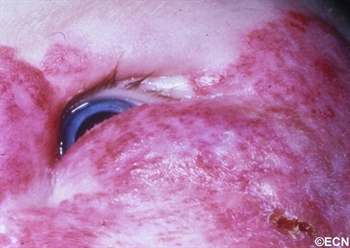
A large periocular hemangioma involves both the upper and lower eyelids. The eye is able to open but there is an astigmatism induced by a mass effect from the lower eye lid.

Children can either be born with or develop reddish “strawberry” colored tumors on or around their eyes. This is one of the most common tumors of infancy. It is 3 times as frequent in girls and can run in families.

Symptoms

Periocular hemangioma of childhood can be large, and commonly grow during the first year of life, but also tend to get smaller (involute) over the following 2 years.

Periocular hemangioma of childhood can extend into the orbit  (behind the eye) and push the eye forward (proptosis), make the eyes misaligned (strabismus), or can cause the eyelid to droop (ptosis).

Children with periocular hemangioma of childhood can have hemangiomas in other parts of their bodies, so a pediatric consultation is necessary. If the hemangiomas are multiple or on the jaw or neck, a pulmonary consultation is necessary to rule out upper respiratory tract involvement. Consider the PHACES syndrome (anomalies of the Posterior fossa, Hemangiomas, the Arteries, Cardiac, Eye, Sternum) which is more commonly seen in girls.

Diagnosis

Hemangioma can be diagnosed by ocular examination with magnetic resonance imaging (MRI). Rarely, a small biopsy may be required to confirm the clinical diagnosis.

Treatments
histopathology-full;size$350,414.ImageHandler
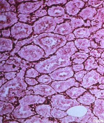
Histopathology reveals multiple well differentiated vascular channels. No atypical cells are seen.

Since periocular hemangioma of childhood is a benign tumor (not a cancer), immediate treatment is often not necessary. In fact, after an initial growth phase, many of these tumors will get smaller by themselves. Most patients can be followed for evidence of spontaneous remission.

Treatment is urgently indicated if the periocular hemangioma of childhood is found to harm the proper development of vision in the affected eye in infants and young children (amblyopia), and for psychosocial reasons in older children and adults.

Let me explain Amblyopia: In order for an eye to achieve its best possible potential for vision, two things are necessary. First, proper images must be focused on the retina and second, the brain must receive those images. During the first 10 years of life, there is a process where images are collected and the brain learns to understand those images. If an eye is blocked by the tumor (or the eye lid), or if the eyes are not aligned, or if the eye is not able to focus images on the retina, the child’s eye-brain connection will not develop. That is, the child will not learn to see from that eye (a problem called amblyopia). Urgent treatment of periocular hemangioma of childhood can be necessary be to prevent amblyopia.

Periocular hemangioma of childhood has been treated with surgery, laser-surgery, radiation, and drugs (intralesional steroids and systemic beta-blockers). When possible, treatment of periocular hemangiomas of childhood involves injections of steroid into the tumor. In comparison to taking the medicine by mouth (PO) or by vein (IV), this approach has the advantage of putting the medicine right into the tumor. An acute effect, but does carry risk of tumor and orbital hemorrhage.

References

  1.  Haik B, Karcioglu Z, Gordon RA, Pechous BP. Capillary hemangioma (infantile periocular hemangioma). Survey of Ophthalmology 1994;38:399-426.
  2.  Kushner BJ. Hemangiomas. Archives of Ophthalmology 2001;118;835-836.

Related Links


About Orbital Tumors: General Information

By Paul T. Finger, MD

Description

Tumors and inflammations can occur behind the eye. They often push the eye forward causing a bulging of the eye called proptosis.

Tumors and inflammations can occur behind and around the eye. They can push the eye forward causing a bulging of the eye called proptosis (image). Alternatively, if the bulging does not allow the eye lids to close, orbital tumor proptosis can cause corneal breakdown. The most common causes of proptosis are thyroid eye disease and lymphoid tumors (lymphoma and atypical lymphoid hyperplasia).

Other tumors include vascular tumors (e.g. hemangiomas, lymphangioma, hemangiopericytoma), lacrimal gland tumors (e.g. dacryoadenitis, benign mixed tumor, sarcoidosis and adenoid cystic carcinoma), and growths that extend from the sinuses into the orbit (e.g. squamous carcinoma, mucocele). Metastatic cancer  can come from other parts of the body to form an orbital tumor. Lastly, an orbit tumor can also be caused by inflammation (e.g. pseudotumor, sarcoidosis) or infection (abscess).

Symptoms

Most patients with orbital tumors notice a bulging of the eyeball or double vision (diplopia). Infections, inflammations and certain orbital cancers can cause pain. Less commonly, orbital tumors are accidentally discovered when patients have a CT or MRI of the head, sinuses and orbit.

Diagnosis

Though CT, MRI’s and ultrasound can help in determining the probable “clinical” diagnosis, most orbital tumors are diagnosed by a surgical biopsy called an orbitotomy (anterior or lateral). During biopsy a specimen is sent to an ophthalmic pathologist who helps determine the exact diagnosis. Dr. Finger has developed a small incision method of orbital biopsy called “FACT.”

Treatments

When possible, orbital tumors are totally removed. If they cannot be removed or if removal will cause too much damage to other important structures around the eye, a piece of tumor may be removed, sent for evaluation by a pathologist and the patient is treated with radiation and/or chemotherapy. Occasionally an orbital tumor is too big or involves the sinuses and requires more extensive surgery with bone-flaps.

If tumors cannot be removed during surgery, most orbital tumors can be treated with external beam radiation therapy. Certain rare orbital tumors require removal of the eye and orbital contents. However, in select cases alternative therapies (e.g. orbital radiotherapy and chemotherapy) can be used to treat residual tumor to spare vision and the eye.

Related links


Patient Stories

"Very well treated by Dr. Finger. He explained everything I needed to know about my issue with detail and attention, putting me at ease and giving me confidence to handle this problem for the rest of my life.”
N.N.

Learn More About Our Patient Care

REQUEST AN APPOINTMENT

Go to Appointment Form

CONNECT AND SHARE

4.5 Stars>
Health Grades
Five Stars>
Rate MDs