Periocular Hemangioma of Childhood

By Paul T. Finger, MD

Description
periocular-hemangioma-full;size$350,248.ImageHandler
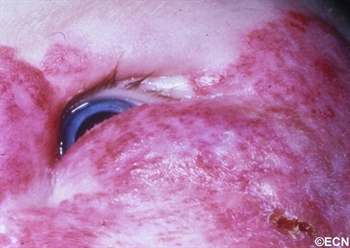
A large periocular hemangioma involves both the upper and lower eyelids. The eye is able to open but there is an astigmatism induced by a mass effect from the lower eye lid.

Children can either be born with or develop reddish “strawberry” colored tumors on or around their eyes. This is one of the most common tumors of infancy. It is 3 times as frequent in girls and can run in families.

Symptoms

Periocular hemangioma of childhood can be large, and commonly grow during the first year of life, but also tend to get smaller (involute) over the following 2 years.

Periocular hemangioma of childhood can extend into the orbit  (behind the eye) and push the eye forward (proptosis), make the eyes misaligned (strabismus), or can cause the eyelid to droop (ptosis).

Children with periocular hemangioma of childhood can have hemangiomas in other parts of their bodies, so a pediatric consultation is necessary. If the hemangiomas are multiple or on the jaw or neck, a pulmonary consultation is necessary to rule out upper respiratory tract involvement. Consider the PHACES syndrome (anomalies of the Posterior fossa, Hemangiomas, the Arteries, Cardiac, Eye, Sternum) which is more commonly seen in girls.

Diagnosis

Hemangioma can be diagnosed by ocular examination with magnetic resonance imaging (MRI). Rarely, a small biopsy may be required to confirm the clinical diagnosis.

Treatments
histopathology-full;size$350,414.ImageHandler
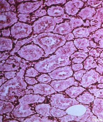
Histopathology reveals multiple well differentiated vascular channels. No atypical cells are seen.

Since periocular hemangioma of childhood is a benign tumor (not a cancer), immediate treatment is often not necessary. In fact, after an initial growth phase, many of these tumors will get smaller by themselves. Most patients can be followed for evidence of spontaneous remission.

Treatment is urgently indicated if the periocular hemangioma of childhood is found to harm the proper development of vision in the affected eye in infants and young children (amblyopia), and for psychosocial reasons in older children and adults.

Let me explain Amblyopia: In order for an eye to achieve its best possible potential for vision, two things are necessary. First, proper images must be focused on the retina and second, the brain must receive those images. During the first 10 years of life, there is a process where images are collected and the brain learns to understand those images. If an eye is blocked by the tumor (or the eye lid), or if the eyes are not aligned, or if the eye is not able to focus images on the retina, the child’s eye-brain connection will not develop. That is, the child will not learn to see from that eye (a problem called amblyopia). Urgent treatment of periocular hemangioma of childhood can be necessary be to prevent amblyopia.

Periocular hemangioma of childhood has been treated with surgery, laser-surgery, radiation, and drugs (intralesional steroids and systemic beta-blockers). When possible, treatment of periocular hemangiomas of childhood involves injections of steroid into the tumor. In comparison to taking the medicine by mouth (PO) or by vein (IV), this approach has the advantage of putting the medicine right into the tumor. An acute effect, but does carry risk of tumor and orbital hemorrhage.

References

  1.  Haik B, Karcioglu Z, Gordon RA, Pechous BP. Capillary hemangioma (infantile periocular hemangioma). Survey of Ophthalmology 1994;38:399-426.
  2.  Kushner BJ. Hemangiomas. Archives of Ophthalmology 2001;118;835-836.

Related Links


Orbital Rhabdomyosarcoma

By Paul T. Finger, MD

Description

orbital_rhabdomyosarcoma
An Orbital Rhabdomyosarcoma

Rhabdomyosarcoma is the most common primary malignancy of the orbit in children. It can also occur in adults, though the average age of patients affected by rhabdomyosarcoma is 7 – 8 years.

Symptoms

Most parents first notice a droopy eyelid (called ptosis), and that the eye is more prominent (called proptosis), or that their child has a tumor under the conjunctival membrane that covers the eye (globe). Rhabdomyosarcoma is usually found in the superonasal orbit (that is under the upper lid near the nose).

orbital_rhabdomyoscaroma_ct
CT of Orbital Rhabdomyosarcoma

Diagnosis

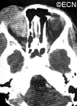
Computed axial tomography (CT-scan) and magnetic resonance imaging (MRI) typically show a mass adjacent to or attached to one of the ocular or orbital muscles. CT is particularly helpful because it offers the best evidence if the orbital bones have been invaded by the rhabdomyosarcoma tumor.

Treatments

Rhabdomyosarcoma can grow rapidly and if the tumor grows into the brain or spreads to the lung, survival is poor. Prompt biopsy of a rhabdomyosarcoma followed by a combination of chemotherapy and irradiation offers the best chance of survival. In fact, recent reports suggest that current treatments offer greater than 90% survival from rhabdomyosarcoma.

Patients will develop problems typically seen after chemotherapy and irradiation of the eye, but if there is no recurrence after 3 years, it is likely that the rhabdomyosarcoma has been controlled.

orbital_rhabdomyosarcoma_histology
Histology of an Orbital Rhabdomyosarcoma

Related Links


Sclerosing Orbital Pseudotumor

By Paul T. Finger, MD

Description

sclerosing_orbital_pseudotumor
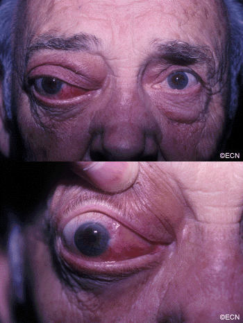
By local growth sclerosing orbital pseudotumors can cause bulging of the eye (proptosis).

Sclerosing orbital pseudotumor is uncommon. Due to unknown reasons, these tumor behave differently than other types of pseudotumor of the orbit. They grow more slowly, cause less pain, and are characterized by scarring (hardening of the tumor tissue). 

Symptoms

Sclerosing orbital pseudotumor is not cancer. But, by local growth it can cause bulging of the eye (proptosis), double vision (diplopia) and loss of vision. Sclerosing orbital pseudotumor can (rarely) extend into the sinuses, brain, and other orbit.

Diagnosis

sclerosing_orbital_pseudotumor_fibrous
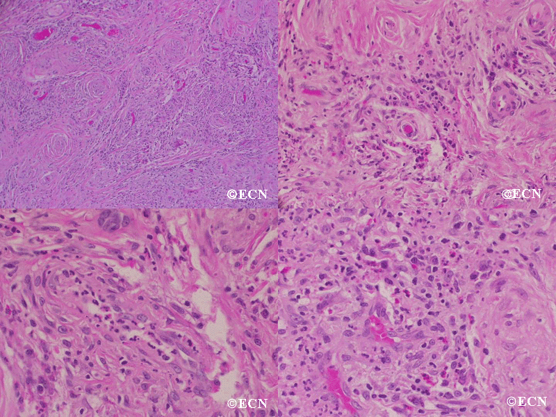
Histopathology reveals large amounts of fibrous tissue.

Sclerosing orbital pseudotumor is usually diagnosed by biospy (orbitotomy).

Once the diagnosis is confirmed by pathology, systemic testing to rule out specific infectious and inflammatory causes should be performed. For example, an ANCA blood test and a chest x-ray should be performed to rule out Wegener’s Granulomatosis. Many of these patients have a past medical history of sinusitis, sinus surgery, or inhalation drug abuse. Therefore, concurrent treatable sinus disease should be addressed.

Treatments

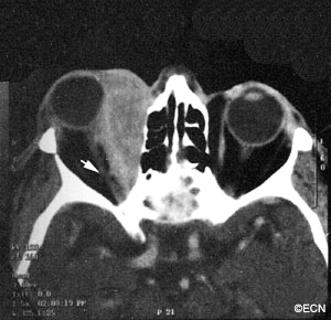
In this case, computed tomographic (CT) scanning demonstrates a mass in the nasal orbit. The arrow demonstrates the optic nerve on stretch. The eye wall (sclera) is indented and the eye pushed out (proptosis).

Sclerosing orbital pseudotumors are less responsive to steroid therapy. Most cases are treated with combinations of surgery, steroid therapy, radiation and chemotherapy depending upon the clinical picture and the patient’s response to treatment.

Related Links


Orbital Pseudotumor

By Paul T. Finger, MD

Description

Inflammations can affect the tissues around the eye (orbit and adnexa). Certain orbital inflammations can look like tumors and are therefore called orbital pseudotumor. Orbital pseudotumor can affect one or both eyes of relatively young patients (less than 50 years old). They are not cancer.

Symptoms

Orbital pseudotumor can be quite painful. In fact, pain is one of the most prominent characteristics of this disease. In addition to pain, an inflammatory mass (tumor) can make the patient’s eye protrude (proptosis) and restrict the movement of the eye. A biopsy (called an orbitotomy) is commonly performed to confirm the diagnosis of orbital pseudotumor and to obtain tissue for pathology examination.

Diagnosis

Orbital pseudotumor is typically characterized by the rapid development of pain, proptosis, and swelling around the eye and orbit in adults. Ultrasound and computed tomographic (CT) scanning typically shows a diffuse infiltration of the orbit, an inflammation of the eye wall (sclera), and/or T-sign (with the optic nerve). Orbital pseudotumor related orbital masses typically have poorly defined margins. Systemic testing (blood and spinal fluid) may show signs of inflammation (e.g. increased sedimentation-rate) or atypical cells.

Patients with classic findings of orbital pseudotumor may be treated without a biopsy. A rapid complete response to steroid therapy helps confirm the diagnosis. Atypical cases of orbital pseudotumor usually undergo a diagnostic biopsy.

Specimens can be sent to test for infectious causes of orbital inflammation and certain systemic diseases. Typically eye cancer specialists will obtain blood, skin and radiographic (e.g. x-ray, MRI) tests for a variety of diseases such as sarcoidosis, tuberculosis, and Wegener’s Granulomatosis (see table below). An orbital biopsy can be particularly helpful in diagnosing many of these disorders.

Treatments

Orbital pseudotumor will respond rapidly to high-dose steroid therapy. Unfortunately, when the steroids are stopped, the inflammation often returns. Eye cancer specialists must reduce the steroid medication very slowly in order to prevent recurrence (return) of the disease.

In certain cases, chemotherapy (e.g. methotrexate, cyclosporine) and low-dose radiation (e.g. 1500-2500 cGy EBRT) may be needed to control the inflammation related to orbital pseudotumor. Most patients do well with steroid therapy but they are always at risk for recurrent orbital pseudotumor.

Additional info

Other Common Causes of Orbital Swelling and Inflammation:

  • Thyroid Eye Disease
  • Sarcoidosis
  • Infectious Orbital Cellulitis
  • Orbital Myositis
  • Scleritis
  • Orbital Vasculitis
  • Sjogren’s Disease
  • Wegener’s Granulomatosis
  • Malignant Ocular Tumors

Related Links


Patient Stories

"Very well treated by Dr. Finger. He explained everything I needed to know about my issue with detail and attention, putting me at ease and giving me confidence to handle this problem for the rest of my life.”
N.N.

Read More

REQUEST AN APPOINTMENT

Go to Appointment Form

CONNECT AND SHARE