By Paul T. Finger, MD
Description
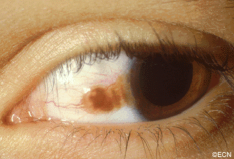
Malignant melanomas can start as a nevus/freckle or arise as newly formed conjunctival pigmentation called primary acquired melanosis (PAM).
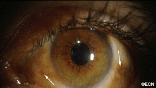
A simple biopsy can determine whether a pigmented conjunctival tumor is a nevus, primary acquired melanosis, or conjunctival melanoma. As seen below, primary acquired melanosis typically affects one eye, in middle-aged, fair-skinned people.
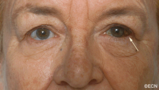
In contrast, darkly pigmented individuals often have naturally occurring pigment on their conjunctiva. When this occurs it is called racial melanosis. Unlike primary acquired melanosis, racial melanosis tends to involve both eyes and is typically present for the entire life of the patient.A simple biopsy can determine whether a pigmented conjunctival tumor is a nevus, primary acquired melanosis, or conjunctival melanoma. As seen above, primary acquired melanosis typically affects one eye, in middle-aged, fair-skinned people.
Symptoms
Pigmentation of the surface of the eye and/or eyelids.
Diagnosis
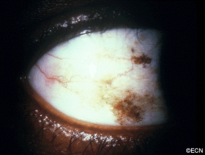
Most pigmented spots on the eye are benign. Your eye care specialist can take a photograph of them and watch to see if they change prior to consideration of biopsy or treatment.
Pigmented conjunctival tumors that are raised, hypervascular, or extend onto the cornea are considered suspicious. Though suspicious conjunctival tumors can be biopsied after your first visit to the eye cancer specialist, close observation for evidence of growth (prior to biopsy) may also be recommended. Documented tumor growth is a strong indicator that biopsy should be performed.
Once the biopsy is performed, the specimen should be evaluated by an ophthalmic pathologist. If there is no ophthalmic pathologist at your center, the slides can be sent for second opinion.
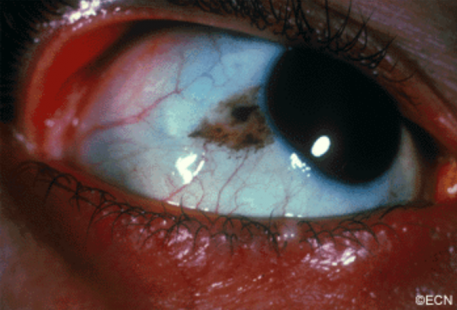
A pigmented conjunctival nevus can be photographed and followed for evidence of growth prior to biopsy or excision. It is important to note that both benign and malignant tumors can grow (though malignant tumors will grow faster).
Slit-lamp photography of benign conjunctival lesions is helpful in determining if subsequent change has occurred. It is a good idea for the patient to have a copy of the initial photograph because doctors are not required to keep medical records indefinitely, and the lesion can change even years after the initial diagnosis.
Related links
Search the Scientific Literature on Pigmented Conjunctival Tumors


