Lymphangioma of the Orbit

Description
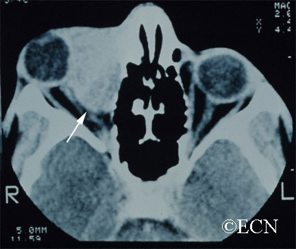
Lymphangioma is rare (less than 7% of childhood orbital tumors). Patients can present with acute proptosis (bulging eye) after minor head trauma, as a gradual proptosis, or after an upper respiratory infection.
Symptoms
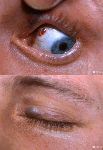
Lymphangioma tends to start in the superior and nasal orbital quadrants. More than 50% affect anterior (conjunctival and adnexal) structures. Typically, the lymphangioma bleeds into itself causing cysts of blood (called chocolate-cysts) within the tumor. If the cyst forms behind the eye, it pushes the eye forward. If the tumor forms in the eyelid or structures around the eye “adnexa”, blood filled lymphatic channels called “lymphangiectasias” can be seen beneath the conjunctiva
Diagnosis

Lymphangioma is usually diagnosed by an eye cancer specialist. A careful history may reveal sudden painful proptosis (bulging of the eye), facial trauma or that the tumor or proptosis started right after a upper respiratory infection.
Severe cases can be associated with corneal exposure, ulceration and optic nerve damage.
Treatments
Though lymphangioma patients can present with a history of sudden proptosis (due to bleeding within the tumor), orbital lymphangiomas are typically slow growing. Therefore, most lymphangiomas are followed by observation for growth-related damage as documented by (clinical and radiographic studies) prior to intervention.
Thus, treatment of lymphangioma is indicated when associated with growth, optic nerve compression, corneal exposure problems (keratitis sicca), glaucoma or vision loss.
When treatment of lymphangioma is considered, the goal is rarely complete removal. This is because the edges of most orbital lymphangiomas are poorly defined. When necessary, patients undergo debulking surgeries with a goal of relieving acute optic nerve compression or corneal exposure. In rare cases, orbital lymphangioma patients may require orbital radiation therapy or exenteration for relief of pain.


