Von Hippel Lindau Disease

By Paul T. Finger, MD

Description

Von Hippel-Lindau (VHL) disease is an autosomal dominantly inherited multisystem cancer syndrome with a predilection for the central nervous system (CNS) and the retina. Retinal capillary “Vvon Hippel” hemangioma is one of the most common and often the earliest manifestations of VHL disease and, therefore, ophthalmologists are frequently involved in the care of patients with this disease. The incidence of VHL disease is approximately one in 40,000 live births and it is estimated that there are approximately 7,000 patients with VHL disease in the United States.

Symptoms

Ophthalmic Findings:

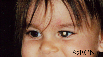
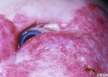
The main ophthalmic finding in VHL is retinal capillary hemangioma, which is a benign hamartoma. The anterior segment can be secondarily involved due to complications such as neovascular glaucoma and cataract formation. A large cohort study found only 2% of eyes had neovascularization of the iris. If a patient has a solitary retinal capillary hemangioma, they do not necessarily have VHL disease. However, they should undergo genetic testing.

Diagnosis

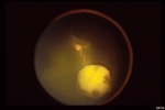
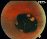
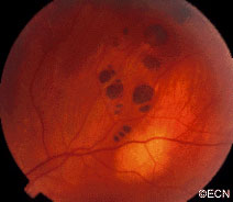
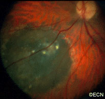
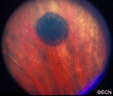
Retinal capillary hemangiomas are usually orange red, circumscribed, round, vascular tumors supplied by a pair of dilated and tortuous feeder vessels. They are most commonly located in the temporal peripheral retina. Juxtapapillary retinal capillary hemangiomas are less common, representing about 11% to 15% of cases, and their appearance can vary depending on whether the lesion is endophytic (grow from the retina into the vitreous gel of the eye), exophytic (grow beneath the retina), or sessile (flat).

Retinal capillary hemangioma usually manifests as a solitary tumor, but approximately one-third of patients have multiple retinal hemangiomas, and up to half of patients have bilateral involvement.

Secondary effects (from the retinal angioma) leading to visual loss, such as intraretinal and subretinal exudation, are often limited to the vicinity of the hemangioma but can be remote, often producing a macular star of exudates. Glial proliferation can lead to tractional retinal detachment and macular pucker. Retinal or vitreal hemorrhages are rarely observed, occurring in fewer than 3% of cases.

Frequency and Patient Related Findings: 

The frequency of occurrence of retinal capillary hemangiomas in VHL disease has been
reported to vary from 49% to 85%. The mean age at diagnosis of retinal capillary hemangioma in VHL disease is approximately 25 years, and most patients present between the ages of 10 and 40 years. The probability of developing a retinal capillary hemangiomas increases progressively with age. Recent publications indicate that the hemangioma is usually manifested by age 30, and the prevalence rate is stable thereafter. Therefore adults with a normal retina at age 30 years may have a low risk of developing a retinal capillary hemangioma during the reminder of their lives. The natural course of retinal capillary hemangiomas is variable (progression, stability or spontaneous regression). Small lesions may remain stable for years or may show evidence of gliosis without leakage, but some have been documented to enlarge. Most hemangiomas, however, tend to enlarge progressively and lead to retinal changes. In late stages they may cause massive exudation and retinal detachment, uveitis (inflammation), glaucoma and phthisis (shrinking of the eye). Classification systems to aid in staging the clinical progression have been developed.

Other Tumors That May Look Like Von Hippel Angiomas: 

The fundus findings of retinal capillary hemangioma are usually typical, and diagnosis can be made based on ophthalmoscopic examination. The diagnosis might be confused with retinal macroaneurysm or adult Coats disease when severe exudation exists.

Treatments

For treatment of Von Hippel retinal angiomas, see Conditions (Von Hippel Angioma) However, in ths section we will discuss the diagnosis of systemic disease and genetics:

The systemic manifestations of VHL are multiple and include CNS hemangiomas of the brain and spinal cord, renal cell carcinomas, renal cysts, pheochromocytomas, pancreatic cysts, islet cell tumors, epididymal cystadenomas, endolymphatic sac tumors of the inner ear, and adnexal papillary cystadenomas of the broad ligament. After retinal capillary hemangioma, the most frequently affected organ systems are the CNS, kidneys and adrenal glands, many of them occurring years after the initial presentation with retinal capillary hemangiomas.

The diagnosis of VHL disease is based on three elements which include: 

  1. retinal capillary hemangioma or CNS hemangioma
  2. visceral lesions
  3. family history of similar lesions

Surveillance: 

After diagnosis is made, screening protocols should be followed, including urinary catecholamines and ophthalmoscopy on an annual basis with MRI of the brain and spinal cord every 2 to 3 years, and yearly abdominal US with an additional abdominal CT scan every 2 to 3 years.

Genetic Testing:

VHL disease is an autosomal dominant disease whose gene is located on chromosome 3p 25-26. The gene functions as a tumor suppressor gene that promotes tumor formation when its function is lost. The normal protein product of the VHL gene forms a complex with other proteins that targets hypoxia inducible factors (HIFs) for degradation. Mutations in the VHL gene result in stabilization of the HIFs, which bind to specific enhancer elements in the VEGF gene and stimulate angiogenesis. With a near-complete penetrance of the disease and only rare instances of mosaicism, genetic testing has been proved helpful in early diagnosis and clinical screening for disease manifestations.

SUMMARY 

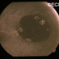
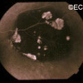
VHL disease is an autosomal dominantly inherited multisystem cancer syndrome with a predilection for the central nervous system and the retina. Retinal capillary hemangioma is one of the most common and earliest manifestations of VHL disease. Fundus findings are usually typical, and diagnosis can be made based on ophthalmoscopic examination, but fluorescein angiography is an additional informative diagnostic tool. Various treatment modalities exist, although the mainstays of therapy are laser photocoagulation and cryotherapy. VHL disease, however, is associated with significant mortality secondary to either CNS hemangioma or renal cell carcinoma. Life expectancy of affected individuals can be improved by early detection, genetic testing and systemic treatment. Treatment of this syndrome requires cooperation of physicians from multiple specialists, including those to treat the central nervous system and kidney tumors.

References

  1. Annesly WJ, Leonard BC, Shields JA, Tasman WS. Fifteen year review of treated cases of retinal angiomatosis. Trans Am Acad Ophthalmol Otolaryngol. 1977;83:446-453.
  2. Choyke PL, Glenn GM, Walther et al. The natural history of renal lesions in von Hippel- Lindau disease: a serial CT study in 28 patients. Am J Roentgenol. 1992;159:1229-1234.
  3. Dahr SS, Cusick M, Roudriguez-Coleman H, et al. Intravitreal anti-vascular endothelial growth factor therapy with pegaptanib for advanced von Hippel-Lindau disease of the retina. Retina. 2007;27:150-158.
  4. Harris AL. von Hippel-Lindau syndrome. Target for anti-vascular endothelial growth factor (VEGF) receptor therapy. The Oncologist. 2000;5(suppl):32-36.
  5. Madhusudan S, Deplanque G, Braybrooke JP, et al. Antiangiogenic therapy for von Hippel-Lindau disease. JAMA. 2004;291:943-944.
  6. Magee MA, Kroll AJ, Lou PL, Ryan EA. Retinal capillary hemangiomas and von Hippel-Lindau disease. Semin Opthalmol. 2006;21:143-150.
  7. Maher ER, Yates JR, Harries, et al. Clinical features and natural history of von Hippel-Lindau disease. QJM. 1990;77:1151-1163.
  8. Maher ER, Lselius L, Yates JR, Et al. von Hippel-Lindau disease: a genetic study. J Med Genet. 1991;28:443-447.
  9. Shields CL, Shields JA, Barret J, et al. Vasoproliferative tumors of the ocular fundus. Classification and clinical manifestations in 103 patients. Arch Ophthalmol. 1995;113:615-623.
  10. Sigelman J. Retinal diseases. Pathogenesis, laser therapy and surgery. Boston:Little Brown and Company. 1984:316.
  11. Singh AD, Shields CL, Shields JA. von Hippel-Lindau disease. Surv Ophthalmol. 2001;46:117-142.
  12. Vail D. Angiomatosis retinae, eleven years after diathermy coagulation. Am J Ophthalmol. 1958;46:525-534.
  13. Webster AR, Maher ER, Moore AT. Clinical characteristics of ocular angiomatosis in von Hippel-Lindau disease and correlation with germline mutation. Arch Ophthalmol. 1999;117:371-378.
  14. Wong WT, Chew EY. Ocular von Hippel-Lindau disease: clinical update and emerging treatments. Curr Opin Ophthalmol. 2008;19:213-217.
  15. Wong WT, Liang KJ, Hammel K, Coleman HR, Chew EY. Intravitreal ranibizumab therapy for retinal capillary hemangioblastoma related to von Hippel-Lindau disease. Ophthalmology. 2008;115:1957-1964.

Von Hippel Angioma

By Paul T. Finger, MD

Description

von_h
von Hippel angioma

Von Hippel angioma can grow within the retina or optic nerve. They characteristically have a “feeding” retinal arteriole and a “draining” retinal vein. Bilateral involvement can be seen in up to 50% of individuals.

Von Hippel angioma are vascular tumors, not cancers and do not metastasize. Twenty percent of patients will be found to have the von Hippel Lindau Syndrome–associated with cerebellar hemangioma, pheochromocytomas, visceral cysts and renal cell carcinomas.

Symptoms

Von Hippel angioma patients either have no symptoms, or become symptomatic due to secondary retinal detachment or rarely neovascular glaucoma. The symptoms of retinal detachment are flashes of light, spots in the vision (floaters), and loss of vision. The symptoms of neovascular glaucoma are eye pain, light sensitivity, vision loss, and headache.

Diagnosis

Some patients with von Hippel angioma will have a family history of this disease. Von Hippel angioma are usually visible by dilated eye examination (ophthalmoscopy). Ultrasound can be used to measure the tumor’s size, and to evaluate for high internal reflectivity. Ophthalmoscopy typically reveals a dilated feeder artery and draining vein. An associated retinal detachment may be seen around the tumor or may be so large as to cover (obscure) an underlying von Hippel angioma.

von_h_2
Fluorescein Angiography – Note the feeder and draining vessels, as well as the bright “light-bulb” appearance.

Fluorescein Angiography: Eye-care specialists perform studies of the blood vessels in the eye with a synthetic organic dye called fluorescein. The dye is injected into the arm and travels to the blood vessels inside the eye. If a tumor is in the eye, we can see specific characteristics of its circulation which can help us differentiate between it and other types of tumors. Von Hippel angiomas have a unique pattern of circulation with a feeder arteriole and a draining vein. Since the tumor extends  from the retina into the eye (vitreous humor), von Hippel angiomas exhibit intense hyperfluorescence, often compared to a “light-bulb.”

Treatments

Von Hippel angiomas can appear in both an autosomal dominant hereditary or sporadic forms. All patients should be given periodic systemic examinations including imaging studies for cerebellar hemangiomas and renal cell carcinoma. Family members should be examined with indirect ophthalmoscopy. Genetic testing is available (see related links below).

The treatment of retinal capillary hemangiomas can be a challenge to the ophthalmologist due to the presence of bilateral multiple tumors and the likelihood of new tumor formation. Despite treatment, up to 25% of cases can have permanent visual loss of acuity less to than 20/40 in one or both eyes. Various treatment modalities including observation, cryotherapy, plaque radiotherapy, and vitreoretinal surgery have been utilized.

Recent advances in the understanding of VHL protein function and tumorigenesis have led to new treatments targeting the biology of the disease, as opposed to ablative or surgical approaches. Molecules upregulated or increased in the context of a VHL mutation, such as vascular endothelial growth factor (VEGF) and platelet-derived growth factor (PDGF), have been targeted in investigational anti-angiogenic therapies, both in systemic manifestations of the disease and in ocular disease.

OBSERVATION AS TREATMENT


Observation is rarely employed due to the tendency of retinal capillary hemangiomas to progress. However, observation only might be chosen in small (Juxtapapillary hemangiomas (those next to the optic nerve disc) are particularly difficult to treat and are initially managed with observation because they can remain stable for years. As a general rule; since these tumors are not cancer and cannot metastasize; treatment should only be undertaken in case of tumor progression or a threat to visual acuity due to the adverse effect of treatment on the optic nerve and major blood vessels.

LASER PHOTOCOAGULATION

Laser photocoagulation is currently used to treat small retinal capillary hemangiomas located in the retina in eyes with clear media. When possible, we first occlude the feeder artery, then (if necessary) to surround/demarcate the posterior 180 degrees of the tumor, lastly and again if needed directly treat the tumor’s surface. Patients should be informed that multiple laser treatment sessions are typically required. Potential complications include retinal detachment, retinal and vitreous hemorrhages.

CRYOTHERAPY


Typical indications for cryotherapy are anterior retinal location of the hemangioma and massive subretinal fluid, which can reduce the laser energy uptake. Double freeze-thaw technique is employed under indirect ophthalmoscopic observation. A 15-year review found that most all hemangiomas under 3.75 mm in diameter successfully responded to cryotherapy.

ANTI-VEGF STRATEGIES


Recent studies have indicated that anti-VEGF strategies may be effective. However, no large clinical trials have been reported.

Additional Info

  1. Wong WT, Liang KJ, Hammel K, Coleman HR, Chew EY. Intravitreal ranibizumab therapy for retinal capillary hemangioblastoma related to von Hippel-Lindau disease. Ophthalmology. 2008;115:1957-1964.
  2. Annesly WJ, Leonard BC, Shields JA, Tasman WS. Fifteen year review of treated cases of retinal angiomatosis. Trans Am Acad Ophthalmol Otolaryngol. 1977;83:446-453.
  3. Madhusudan S, Deplanque G, Braybrooke JP, et al. Antiangiogenic therapy for von Hippel-Lindau disease. JAMA. 2004;291:943-944.

Retinoblastoma

By Paul T. Finger, MD

Description

retinoblastoma
White pupil “leukocoria” sign of retinoblastoma

Retinoblastoma is the most common intraocular cancer of childhood and affects approximately 300 children in the United States each year. More than 96% of children in North America and Europe are cured of retinoblastoma due to early detection and treatment of the affected eye. This is not true for children in countries that do not have eye cancer specialists.

Unfortunately, some children can have both eyes affected. Whenever possible, eye-cancer specialists try to save a child’s eye and preserve their vision.

Symptoms

retinoblastoma_endo-full;size$150,100.ImageHandler
An intraocular photograph of an isolated “endophytic” retinoblastoma.

Leukocoria (white pupil) and misaligned eyes (strabismus) are the most common signs of retinoblastoma. In other cases, the child may have developed neovascular glaucoma and may be in pain. Longstanding glaucoma can cause enlargement of the eye (buphthalmos). Children with neovascular glaucoma and enlargement of the eye are at greater risk for extraocular spread of their retinoblastoma.

A family history of retinoblastoma can be very important. Retinoblastoma was the first cancer to be directly associated with a genetic abnormality (Deletions or mutation of the q14 band of chromosome 13). Retinoblastoma can occur sporadically (without a family history) or it can be inherited (with a family history).

If a genetic mutation is found there is a 45-50% chance that the parents will have another child with retinoblastoma. If there is no family history and no mutation is found, the risk of having a second child with retinoblastoma is 2-5%. The average age of children first diagnosed with retinoblastoma is 18 months (typical range 0 to 36 months).

Diagnosis

More than 75% of children with retinoblastoma are first noted to have a “white-pupil” (which the doctors call leukocoria), or poorly aligned eyes (which the doctors call strabismus), or a red and painful eye (usually due to glaucoma). Other eye diseases which can cause these symptoms include congenital cataract, Toxocara canis, Coat’s disease, and persistent hypertrophic primary vitreous (PHPV). These diseases may look like retinoblastoma, but by performing an eye examination under anesthesia (EUA), specialized blood tests, digital photography, radiographic scans, and ultrasound evaluations ophthalmic oncologists can diagnose intraocular retinoblastoma in over 95% of cases. In order to be 100% correct all the time, eye-cancer specialists would have to perform a biopsy. Biopsies of intraocular retinoblastoma are avoided in order to prevent cancer cells from spreading outside of the eye.

The presence of orbital extension, uveal involvement, and optic nerve invasion are known risk factors for the development of metastatic retinoblastoma.

Treatments

Retinoblastoma treatment typically requires the cooperation of an ophthalmic oncologist, pediatric oncologist, and radiation therapist. Over the last 30 years, treatment has evolved from simple enucleation (removal of the eye), to eye-sparing radiotherapy, and more recently to chemotherapy-based multi-modality therapy (for selected cases). Intra-arterial chemotherapy (IAC) has recently been investigated to save eyes, vision and spare the child from systemic chemotherapy.

Though retinoblastoma has been cured by external beam irradiation, investigators have found that radiation may cause an increase in the risk of developing second cancers later in life.

Protocols are currently being evaluated to use chemotherapy to shrink the retinoblastoma in order to treat them with laser therapy, freezing therapy (cryotherapy), and local “plaque” radiation. Where applicable, these techniques are thought to be safer than external beam irradiation for retinoblastoma. Intra-arterial chemotherapy is a newer method of perfusing the eye with chemotherapy, used for selected cases.

Treatment of retinoblastoma often requires a team of doctors made up of ophthalmic, radiation and pediatric oncologists. These doctors should evaluate your child, discuss all the different forms of treatment, and make them available.

Related links


Retinal Pigment Epithelium (RPE) Tumors

By Paul T. Finger, MD

Description

Adenocarcinomas and adenomas typically arise from a part of the retina called the retinal pigment epithelium or RPE. They can also occur in the ciliary body. Though all are rare, benign adenomas are much more common than RPE cancers. Less than 20 malignant adenocarcinomas of the RPE have ever been reported.  

Further, during the modern era of reporting, malignant adenocarcinomas of the RPE have never been reported to spread to other parts of the body.

In the past, almost all RPE adenocarcinomas were clinically diagnosed as atypical choroidal melanomas and the eyes were enucleated. In Dr. Finger’s review of the literature, RPE adenocarcinomas were found to be more common in females, more likely to cause intraocular inflammation (atypical for choroidal melanomas) and have responded poorly to radiation therapy.

Symptoms

Patients with adenomas of the retinal pigment epithelium typically have no symptoms. These tumors are found during routine eye examination and referred to eye tumor specialists for further evaluation. 

Diagnosis

rpe_tumors
Ultrasound of an RPE adenocarcinoma reveals a “stuck on” the choroid appearance.

Retinal adenoma and adenocarcinoma will appear black. They originate from the retina, so they are not likely to have a surrounding or overlying retinal detachment. Adenomas appear dark. “block fluorescence” on fluorescein angiography.

Ultrasound may be helpful in distinguishing adenoma and adenocarcinoma of the RPE from uveal melanomas. Consider that they tend to arise from the retina above Bruch’s membrane. Therefore, they appear to be stuck on rather than growing from the underlying choroid. Several investigators have noted a tuberous rather than collar-button or dome-shape.

Treatments

Retinal adenoma should be photographed, measured by ultrasound and followed for evidence of growth prior to consideration of treatment.

rpe_tumors_3
Histopathologic evaluation of the same tumor shows that it arises from the retina.

In the rare instance of retinal adenocarcinoma, most patients have been enucleated with a presumed diagnosis of choroidal melanoma. Should adenocarcinoma of the retinal pigment epithelium be suspected, a biopsy should be considered. Characteristics of RPE adenocarcinoma are a female patient with a dark intraocular tumor and associated with iritis and/or uveitis.


Retinal Pigment Epithelial (RPE) Hypertrophy

By Paul T. Finger, MD

Description

rpe_black_well
Note the flat, black well circumscribed lesion with areas of retinal pigment epithelial atrophy.

The retinal pigment epithelium (RPE) is a pigmented layer of the retina which can be thicker than normal at birth (congenital) or may thicken later in life. Areas of retinal pigment epithelial (RPE) hypertrophy usually do not cause symptoms. They are typically found during routine eye examinations.

Congenital retinal pigment epithelial hypertrophy (CHRPE) is usually found before patients reach 30 years of age. They may enlarge with time, but are not malignant. CHPRE has been an association with Gardner’s Syndrome (familial colonic polyposis).

congenital hypertrophy of the retinal pigment epithelium, "bear-tracks."
This is a case of congenital hypertrophy of the retinal pigment epithelium, “bear-tracks.”

Therefore, if your eye care specialist has told you that you have CHPRE( pronounced CHER PEE), it is reasonable to tell your primary care physician or gastroenterologist (if you have one) so that he or she may recommend the frequency of colon screening tests.

In contrast, acquired retinal pigment epithelial hypertrophy (RPEH) is typically found later in life. They are typically jet-black to gray, flat, with a halo around its edges. Variable in size, RPEH lesions may develop lacunae of lightly colored areas of atrophy (see image above). These

blue hued retinal pigment epithelial hypertrophy
This area of retinal pigment epithelial hypertrophy demonstrates a blue hue.

tumors are more commonly found in the peripheral retina where thickness is more difficult to judge by ophthalmoscopy.

Symptoms

Almost all patients with retinal pigment epithelial hypertrophy do not have symptoms. These pigmented intraocular lesions are found by eye care specialists during dilated examination of the inside of the eye (ophthalmoscopy). Eye tumor specialists can typically differentiate between retinal pigment epithelial hypertrophy and melanoma by clinical examination (without a biopsy).

Diagnosis

A small area of retinal pigment epithelial hypertrophy
A small area of retinal pigment epithelial hypertrophy. It appears well circumscribed and with areas of relative lucency at the edges.

Retinal pigment epithelial hypertrophy (CHRPE and RPEH) can be diagnosed by ophthalmic examination. The eye examination will concentrate on the appearance of the retinal pigment epithelial hypertrophy. RPEH lesions tend to be black or atrophic. They may be surrounded by a halo of less pigmented tissue or exhibit a sharp demarcation line.

Ultrasonography typically shows that RPE hypertrophy is flat to minimally elevated and slightly hyper-reflective.

Fluorescein angiography of RPE hypertrophy typically demonstrates blockage of fluorescence (except in the areas of atrophy which are hyperfluorescent).

Optical coherence tomography (OCT) of RPE hypertrophy will demonstrate both thickening and thinning. The overlying retina is thinned, the retinal pigment epithelium is both thickened or can be thinned. The underlying choroid is typically thinned.

Treatment

Photographic documentation of these lesions is recommended for future comparison. Ultrasonography and fluorescein angiography is typically used to differentiate RPE hypertrophy from uveal melanoma and certain rare intraocular tumors. Serial observation is warranted in that RPE hypertrophy can enlarge over time.

Related Links


Periocular Hemangioma of Childhood

By Paul T. Finger, MD

Description

periocular-hemangioma-full;size$350,248.ImageHandler
A large periocular hemangioma involves both the upper and lower eyelids. The eye is able to open but there is an astigmatism induced by a mass effect from the lower eye lid.

Children can either be born with or develop reddish “strawberry” colored tumors on or around their eyes. This is one of the most common tumors of infancy. It is 3 times as frequent in girls and can run in families.

Symptoms

Periocular hemangioma of childhood can be large, and commonly grow during the first year of life, but also tend to get smaller (involute) over the following 2 years.

Periocular hemangioma of childhood can extend into the orbit  (behind the eye) and push the eye forward (proptosis), make the eyes misaligned (strabismus), or can cause the eyelid to droop (ptosis).

Children with periocular hemangioma of childhood can have hemangiomas in other parts of their bodies, so a pediatric consultation is necessary. If the hemangiomas are multiple or on the jaw or neck, a pulmonary consultation is necessary to rule out upper respiratory tract involvement. Consider the PHACES syndrome (anomalies of the Posterior fossa, Hemangiomas, the Arteries, Cardiac, Eye, Sternum) which is more commonly seen in girls.

Diagnosis

Hemangioma can be diagnosed by ocular examination with magnetic resonance imaging (MRI). Rarely, a small biopsy may be required to confirm the clinical diagnosis.

Treatments

histopathology-full;size$350,414.ImageHandler
Histopathology reveals multiple well differentiated vascular channels. No atypical cells are seen.

Since periocular hemangioma of childhood is a benign tumor (not a cancer), immediate treatment is often not necessary. In fact, after an initial growth phase, many of these tumors will get smaller by themselves. Most patients can be followed for evidence of spontaneous remission.

Treatment is urgently indicated if the periocular hemangioma of childhood is found to harm the proper development of vision in the affected eye in infants and young children (amblyopia), and for psychosocial reasons in older children and adults.

Let me explain Amblyopia: In order for an eye to achieve its best possible potential for vision, two things are necessary. First, proper images must be focused on the retina and second, the brain must receive those images. During the first 10 years of life, there is a process where images are collected and the brain learns to understand those images. If an eye is blocked by the tumor (or the eye lid), or if the eyes are not aligned, or if the eye is not able to focus images on the retina, the child’s eye-brain connection will not develop. That is, the child will not learn to see from that eye (a problem called amblyopia). Urgent treatment of periocular hemangioma of childhood can be necessary be to prevent amblyopia.

Periocular hemangioma of childhood has been treated with surgery, laser-surgery, radiation, and drugs (intralesional steroids and systemic beta-blockers). When possible, treatment of periocular hemangiomas of childhood involves injections of steroid into the tumor. In comparison to taking the medicine by mouth (PO) or by vein (IV), this approach has the advantage of putting the medicine right into the tumor. An acute effect, but does carry risk of tumor and orbital hemorrhage.

References

  1.  Haik B, Karcioglu Z, Gordon RA, Pechous BP. Capillary hemangioma (infantile periocular hemangioma). Survey of Ophthalmology 1994;38:399-426.
  2.  Kushner BJ. Hemangiomas. Archives of Ophthalmology 2001;118;835-836.

Related Links


Orbital Rhabdomyosarcoma

By Paul T. Finger, MD

Description

orbital_rhabdomyosarcoma
An Orbital Rhabdomyosarcoma

Rhabdomyosarcoma is the most common primary malignancy of the orbit in children. It can also occur in adults, though the average age of patients affected by rhabdomyosarcoma is 7 – 8 years.

Symptoms

Most parents first notice a droopy eyelid (called ptosis), and that the eye is more prominent (called proptosis), or that their child has a tumor under the conjunctival membrane that covers the eye (globe). Rhabdomyosarcoma is usually found in the superonasal orbit (that is under the upper lid near the nose).

orbital_rhabdomyoscaroma_ct
CT of Orbital Rhabdomyosarcoma

Diagnosis

Computed axial tomography (CT-scan) and magnetic resonance imaging (MRI) typically show a mass adjacent to or attached to one of the ocular or orbital muscles. CT is particularly helpful because it offers the best evidence if the orbital bones have been invaded by the rhabdomyosarcoma tumor.

Treatments

Rhabdomyosarcoma can grow rapidly and if the tumor grows into the brain or spreads to the lung, survival is poor. Prompt biopsy of a rhabdomyosarcoma followed by a combination of chemotherapy and irradiation offers the best chance of survival. In fact, recent reports suggest that current treatments offer greater than 90% survival from rhabdomyosarcoma.

Patients will develop problems typically seen after chemotherapy and irradiation of the eye, but if there is no recurrence after 3 years, it is likely that the rhabdomyosarcoma has been controlled.

orbital_rhabdomyosarcoma_histology
Histology of an Orbital Rhabdomyosarcoma

Related Links


Sclerosing Orbital Pseudotumor

By Paul T. Finger, MD

Description

sclerosing_orbital_pseudotumor
By local growth sclerosing orbital pseudotumors can cause bulging of the eye (proptosis).

Sclerosing orbital pseudotumor is uncommon. Due to unknown reasons, these tumor behave differently than other types of pseudotumor of the orbit. They grow more slowly, cause less pain, and are characterized by scarring (hardening of the tumor tissue). 

Symptoms

Sclerosing orbital pseudotumor is not cancer. But, by local growth it can cause bulging of the eye (proptosis), double vision (diplopia) and loss of vision. Sclerosing orbital pseudotumor can (rarely) extend into the sinuses, brain, and other orbit.

Diagnosis

sclerosing_orbital_pseudotumor_fibrous
Histopathology reveals large amounts of fibrous tissue.

Sclerosing orbital pseudotumor is usually diagnosed by biospy (orbitotomy).

Once the diagnosis is confirmed by pathology, systemic testing to rule out specific infectious and inflammatory causes should be performed. For example, an ANCA blood test and a chest x-ray should be performed to rule out Wegener’s Granulomatosis. Many of these patients have a past medical history of sinusitis, sinus surgery, or inhalation drug abuse. Therefore, concurrent treatable sinus disease should be addressed.

Treatments

In this case, computed tomographic (CT) scanning demonstrates a mass in the nasal orbit. The arrow demonstrates the optic nerve on stretch. The eye wall (sclera) is indented and the eye pushed out (proptosis).

Sclerosing orbital pseudotumors are less responsive to steroid therapy. Most cases are treated with combinations of surgery, steroid therapy, radiation and chemotherapy depending upon the clinical picture and the patient’s response to treatment.

Related Links


Orbital Pseudotumor

By Paul T. Finger, MD

Description

Inflammations can affect the tissues around the eye (orbit and adnexa). Certain orbital inflammations can look like tumors and are therefore called orbital pseudotumor. Orbital pseudotumor can affect one or both eyes of relatively young patients (less than 50 years old). They are not cancer.

Symptoms

Orbital pseudotumor can be quite painful. In fact, pain is one of the most prominent characteristics of this disease. In addition to pain, an inflammatory mass (tumor) can make the patient’s eye protrude (proptosis) and restrict the movement of the eye. A biopsy (called an orbitotomy) is commonly performed to confirm the diagnosis of orbital pseudotumor and to obtain tissue for pathology examination.

Diagnosis

Orbital pseudotumor is typically characterized by the rapid development of pain, proptosis, and swelling around the eye and orbit in adults. Ultrasound and computed tomographic (CT) scanning typically shows a diffuse infiltration of the orbit, an inflammation of the eye wall (sclera), and/or T-sign (with the optic nerve). Orbital pseudotumor related orbital masses typically have poorly defined margins. Systemic testing (blood and spinal fluid) may show signs of inflammation (e.g. increased sedimentation-rate) or atypical cells.

Patients with classic findings of orbital pseudotumor may be treated without a biopsy. A rapid complete response to steroid therapy helps confirm the diagnosis. Atypical cases of orbital pseudotumor usually undergo a diagnostic biopsy.

Specimens can be sent to test for infectious causes of orbital inflammation and certain systemic diseases. Typically eye cancer specialists will obtain blood, skin and radiographic (e.g. x-ray, MRI) tests for a variety of diseases such as sarcoidosis, tuberculosis, and Wegener’s Granulomatosis (see table below). An orbital biopsy can be particularly helpful in diagnosing many of these disorders.

Treatments

Orbital pseudotumor will respond rapidly to high-dose steroid therapy. Unfortunately, when the steroids are stopped, the inflammation often returns. Eye cancer specialists must reduce the steroid medication very slowly in order to prevent recurrence (return) of the disease.

In certain cases, chemotherapy (e.g. methotrexate, cyclosporine) and low-dose radiation (e.g. 1500-2500 cGy EBRT) may be needed to control the inflammation related to orbital pseudotumor. Most patients do well with steroid therapy but they are always at risk for recurrent orbital pseudotumor.

Additional info

Other Common Causes of Orbital Swelling and Inflammation:

  • Thyroid Eye Disease
  • Sarcoidosis
  • Infectious Orbital Cellulitis
  • Orbital Myositis
  • Scleritis
  • Orbital Vasculitis
  • Sjogren’s Disease
  • Wegener’s Granulomatosis
  • Malignant Ocular Tumors

Related Links


Orbital Mucocele

By Paul T. Finger, MD

Description

aadenoid_cystic
These magnetic resonance imaging (MRI) studies demonstrate displacement of the optic nerve, a bright T1 tumor image, and a variably bright T2 tumor image. The tumor is noted to involve the orbit, the ethmoid and frontal sinuses.

Orbital mucocele can occur when sinus mucoceles cannot naturally drain through the nose. Instead, they grow and slowly invade adjacent orbital tissues.

Generally arising from the ethmoid or frontal sinuses, orbital mucoceles are mucous or fluid filled cysts which can displace the eye. Frontal sinus mucoceles can force the eye down, ethmoid tumors will push the eye out and maxillary lesions can elevate or push the eye in. Lastly, orbital mucoceles originating in the sphenoid sinus can compress the optic nerve resulting in loss of vision.

Symptoms

Patients with mucocele of the orbit, typically have a history of facial trauma or chronic sinus disease. They tend to be in their mid 40’s or older. They can have painless proptosis, or complain of headaches, double vision, or loss of vision.

Diagnosis

orbital_mucocele_axial
Computed axial tomography demonstrates displacement of the medial rectus muscle (MR), as well as erosion and obliteration of portions of the orbital roof (black arrows). Note that the orbital portion of the mucocele is partially encased in bone. This is characteristic of mucocele. The rectus muscles and optic nerve are labeled.

demonstrates displacement of the medial rectus muscle (MR), as well as erosion and obliteration of portions of the orbital roof (black arrows). Note that the orbital portion of the mucocele is partially encased in bone. This is characteristic of mucocele. The rectus muscles and optic nerve are labeled.

Though magnetic resonance imaging (MRI) can be consistent with a mucous or serous fluid-filled tumor, a drainage procedure is typically required and found to be diagnostic. The mucoid or serous fluid (which is found to make up the mucocele) should be sent for culture and sensitivity as well as cytologic examination. Mucoceles can be infected. In those cases, the choice of antibiotics can depend on cultures taken during surgery.

Treatments

orbial_mucocele_transnasal
Intraoperative transnasal video photography is presented to show the mucocele cavity (arrow) after ethmoidectomy (photograph courtesy of Steven Schaefer, MD)

The treament of mucocele of the orbit is surgical. It is best to have a combination of an ophthalmic and ENT surgeons. Treatment involves removal of as much of the cyst and its lining as possible. This usually requires an orbitotomy and sinusectomy. It is most important to re-establish or create a new drainage pathway for the mucous to exit the nose.

Additional info

Case Example: A 63 year old male was referred to The New York Eye Cancer Center with a 6 month history of progressive painless proptosis of the right eye.

Despite this large orbital tumor with optic nerve displacement, the patient was 20/20 OU, he had no visual field defect, and no signs of optic neuropathy. A complete medical survey was initiated and the patient was cleared for surgery. A combination of anterior orbitotomy and transnasal ethmoidectomy were performed to evacuate the mucous and allow for future drainage.

Related links


Patient Stories

"Very well treated by Dr. Finger. He explained everything I needed to know about my issue with detail and attention, putting me at ease and giving me confidence to handle this problem for the rest of my life.”
N.N.

Read More

REQUEST AN APPOINTMENT

Go to Appointment Form

CONNECT AND SHARE