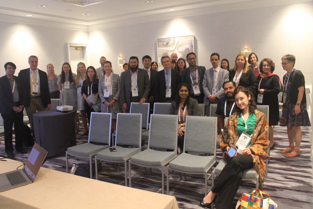
Day 4: Live From the ISOO 2019!

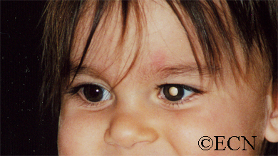
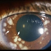
Day 4 at the ISOO: Retinoblastoma

Every two years, eye cancer specialists worldwide convene at the International Society of Ocular Oncology Conference in order to review latest research and encourage international collaboration within the field of ophthalmic oncology.

For details on the ISOO Biennial Conference, read more here.


Preventing Retinoblastoma

Dr. Helen Dimaras opened with her talk on clinically preventable retinoblastoma, having analyzed the frequency of unfavorable outcomes such as death, metastasis, enucleation, EBRT, and how we can avoid such outcomes. Looking at data retrospectively allows physicians to “review how patients do and improve practice by looking at the past, reducing errors, and improving documentation and precision of care.”

In high-income nations, frequency of poor outcomes is low, and we need to work on expanding such rates abroad. Not only this, but a comment from the audience emphasized the significance of this study’s call to action: even just one child who succumbs to retinoblastoma is a child lost, and such outcomes need to be taken very seriously. We need to avoid misdiagnosis and under-treatment as much we humanly can to ensure that unfavorable outcomes do not happen in the future.
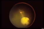
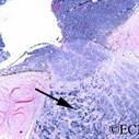

“Chemoplaques”

Dr. Brenda Gallie presented an interesting study regarding a device named “chemoplaques” in Chemoplaque (sustained-release topotecan episcleral device) for retinoblastoma: Opportunity for rapid clinical evaluation of toxicity and efficacy to support safe eye salvage. The devise works through continuous exposure via diffusion, and has thus far produced promising results. By day 28, most of the studied tumors had gone away, and many were gone even earlier, by day 18.


International Fellowship Training Guidelines

Forming Plans for Future Fellowships

In an auxiliary lecture hall, many of us gathered to discuss training fellows all over the world in order to create and develop international treatment centers that will allow greater access to eye cancer care in underdeveloped countries and underserved areas. Sound familiar? As this is one of the primary missions of The Eye Cancer Foundation, Chairman of the ECF, Dr. Finger gave a lecture and lead the discussion.

Dr. Finger with fellows trained by Dr. Honavar in Hyderabad, India. From left: Dr. Sumeet Lahane, Dr. Paul Finger, Dr. Puneet Jain, Dr. Mrithika Sen, Dr. Ankit Tomar, and Dr.

So what is the plan of action? Flexible fellowships with candidates sent from the ophthalmic society of the unserved or underserved region. Being underserved locations, we must be open to 3-month long fellowships, since the physician is needed in their country to give care. 12-month fellowships oftentimes are unfeasible. We must also make a collaborative effort to increase funding for such programs. Dr. Finger says “It has been my experience that funding can come from unexpected places. All you need to do is discuss this wonderful work we are doing and relate how we need help. It is a blessing to allow others to share in the accomplishment of saving children’s sight and life.”

Full video of Dr. Finger’s Lecture Here!

International Fellowship Training Guidelines Meeting Attendees


Retinoblastoma

By Paul T. Finger, MD

Description

retinoblastoma
White pupil “leukocoria” sign of retinoblastoma

Retinoblastoma is the most common intraocular cancer of childhood and affects approximately 300 children in the United States each year. More than 96% of children in North America and Europe are cured of retinoblastoma due to early detection and treatment of the affected eye. This is not true for children in countries that do not have eye cancer specialists.

Unfortunately, some children can have both eyes affected. Whenever possible, eye-cancer specialists try to save a child’s eye and preserve their vision.

Symptoms

retinoblastoma_endo-full;size$150,100.ImageHandler
An intraocular photograph of an isolated “endophytic” retinoblastoma.

Leukocoria (white pupil) and misaligned eyes (strabismus) are the most common signs of retinoblastoma. In other cases, the child may have developed neovascular glaucoma and may be in pain. Longstanding glaucoma can cause enlargement of the eye (buphthalmos). Children with neovascular glaucoma and enlargement of the eye are at greater risk for extraocular spread of their retinoblastoma.

A family history of retinoblastoma can be very important. Retinoblastoma was the first cancer to be directly associated with a genetic abnormality (Deletions or mutation of the q14 band of chromosome 13). Retinoblastoma can occur sporadically (without a family history) or it can be inherited (with a family history).

If a genetic mutation is found there is a 45-50% chance that the parents will have another child with retinoblastoma. If there is no family history and no mutation is found, the risk of having a second child with retinoblastoma is 2-5%. The average age of children first diagnosed with retinoblastoma is 18 months (typical range 0 to 36 months).

Diagnosis

More than 75% of children with retinoblastoma are first noted to have a “white-pupil” (which the doctors call leukocoria), or poorly aligned eyes (which the doctors call strabismus), or a red and painful eye (usually due to glaucoma). Other eye diseases which can cause these symptoms include congenital cataract, Toxocara canis, Coat’s disease, and persistent hypertrophic primary vitreous (PHPV). These diseases may look like retinoblastoma, but by performing an eye examination under anesthesia (EUA), specialized blood tests, digital photography, radiographic scans, and ultrasound evaluations ophthalmic oncologists can diagnose intraocular retinoblastoma in over 95% of cases. In order to be 100% correct all the time, eye-cancer specialists would have to perform a biopsy. Biopsies of intraocular retinoblastoma are avoided in order to prevent cancer cells from spreading outside of the eye.

The presence of orbital extension, uveal involvement, and optic nerve invasion are known risk factors for the development of metastatic retinoblastoma.

Treatments

Retinoblastoma treatment typically requires the cooperation of an ophthalmic oncologist, pediatric oncologist, and radiation therapist. Over the last 30 years, treatment has evolved from simple enucleation (removal of the eye), to eye-sparing radiotherapy, and more recently to chemotherapy-based multi-modality therapy (for selected cases). Intra-arterial chemotherapy (IAC) has recently been investigated to save eyes, vision and spare the child from systemic chemotherapy.

Though retinoblastoma has been cured by external beam irradiation, investigators have found that radiation may cause an increase in the risk of developing second cancers later in life.

Protocols are currently being evaluated to use chemotherapy to shrink the retinoblastoma in order to treat them with laser therapy, freezing therapy (cryotherapy), and local “plaque” radiation. Where applicable, these techniques are thought to be safer than external beam irradiation for retinoblastoma. Intra-arterial chemotherapy is a newer method of perfusing the eye with chemotherapy, used for selected cases.

Treatment of retinoblastoma often requires a team of doctors made up of ophthalmic, radiation and pediatric oncologists. These doctors should evaluate your child, discuss all the different forms of treatment, and make them available.

Related links


Patient Stories

"Very well treated by Dr. Finger. He explained everything I needed to know about my issue with detail and attention, putting me at ease and giving me confidence to handle this problem for the rest of my life.”
N.N.

Read More

REQUEST AN APPOINTMENT

Go to Appointment Form

CONNECT AND SHARE