Retinal Pigment Epithelial (RPE) Hypertrophy
By Paul T. Finger, MD
Description
The retinal pigment epithelium (RPE) is a pigmented layer of the retina which can be thicker than normal at birth (congenital) or may thicken later in life. Areas of retinal pigment epithelial (RPE) hypertrophy usually do not cause symptoms. They are typically found during routine eye examinations.
Congenital retinal pigment epithelial hypertrophy (CHRPE) is usually found before patients reach 30 years of age. They may enlarge with time, but are not malignant. CHPRE has been an association with Gardner’s Syndrome (familial colonic polyposis).
Therefore, if your eye care specialist has told you that you have CHPRE( pronounced CHER PEE), it is reasonable to tell your primary care physician or gastroenterologist (if you have one) so that he or she may recommend the frequency of colon screening tests.
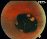
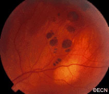
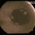
In contrast, acquired retinal pigment epithelial hypertrophy (RPEH) is typically found later in life. They are typically jet-black to gray, flat, with a halo around its edges. Variable in size, RPEH lesions may develop lacunae of lightly colored areas of atrophy (see image above). These
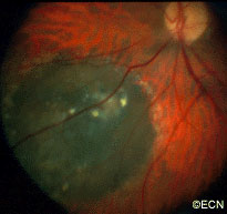
tumors are more commonly found in the peripheral retina where thickness is more difficult to judge by ophthalmoscopy.
Symptoms
Almost all patients with retinal pigment epithelial hypertrophy do not have symptoms. These pigmented intraocular lesions are found by eye care specialists during dilated examination of the inside of the eye (ophthalmoscopy). Eye tumor specialists can typically differentiate between retinal pigment epithelial hypertrophy and melanoma by clinical examination (without a biopsy).
Diagnosis
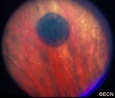
Retinal pigment epithelial hypertrophy (CHRPE and RPEH) can be diagnosed by ophthalmic examination. The eye examination will concentrate on the appearance of the retinal pigment epithelial hypertrophy. RPEH lesions tend to be black or atrophic. They may be surrounded by a halo of less pigmented tissue or exhibit a sharp demarcation line.
Ultrasonography typically shows that RPE hypertrophy is flat to minimally elevated and slightly hyper-reflective.
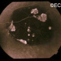
Fluorescein angiography of RPE hypertrophy typically demonstrates blockage of fluorescence (except in the areas of atrophy which are hyperfluorescent).
Optical coherence tomography (OCT) of RPE hypertrophy will demonstrate both thickening and thinning. The overlying retina is thinned, the retinal pigment epithelium is both thickened or can be thinned. The underlying choroid is typically thinned.
Treatment
Photographic documentation of these lesions is recommended for future comparison. Ultrasonography and fluorescein angiography is typically used to differentiate RPE hypertrophy from uveal melanoma and certain rare intraocular tumors. Serial observation is warranted in that RPE hypertrophy can enlarge over time.
Related Links
- Search PubMed for articles on Retinal Pigment Epithelial (RPE) Hypertrophy
- Search Google for information on Retinal Pigment Epithelial (RPE) Hypertrophy


