Periocular Hemangioma of Childhood

By Paul T. Finger, MD

Description

periocular-hemangioma-full;size$350,248.ImageHandler
A large periocular hemangioma involves both the upper and lower eyelids. The eye is able to open but there is an astigmatism induced by a mass effect from the lower eye lid.

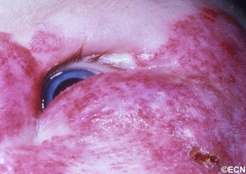
Children can either be born with or develop reddish “strawberry” colored tumors on or around their eyes. This is one of the most common tumors of infancy. It is 3 times as frequent in girls and can run in families.

Symptoms

Periocular hemangioma of childhood can be large, and commonly grow during the first year of life, but also tend to get smaller (involute) over the following 2 years.

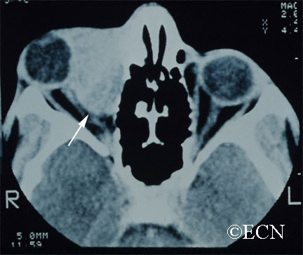
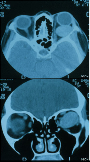
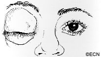
Periocular hemangioma of childhood can extend into the orbit  (behind the eye) and push the eye forward (proptosis), make the eyes misaligned (strabismus), or can cause the eyelid to droop (ptosis).

Children with periocular hemangioma of childhood can have hemangiomas in other parts of their bodies, so a pediatric consultation is necessary. If the hemangiomas are multiple or on the jaw or neck, a pulmonary consultation is necessary to rule out upper respiratory tract involvement. Consider the PHACES syndrome (anomalies of the Posterior fossa, Hemangiomas, the Arteries, Cardiac, Eye, Sternum) which is more commonly seen in girls.
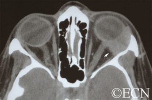
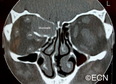
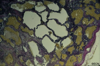
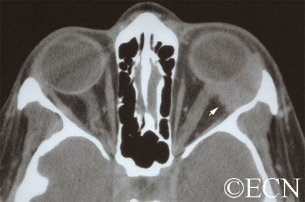
Diagnosis

Hemangioma can be diagnosed by ocular examination with magnetic resonance imaging (MRI). Rarely, a small biopsy may be required to confirm the clinical diagnosis.

Treatments

histopathology-full;size$350,414.ImageHandler
Histopathology reveals multiple well differentiated vascular channels. No atypical cells are seen.

Since periocular hemangioma of childhood is a benign tumor (not a cancer), immediate treatment is often not necessary. In fact, after an initial growth phase, many of these tumors will get smaller by themselves. Most patients can be followed for evidence of spontaneous remission.

Treatment is urgently indicated if the periocular hemangioma of childhood is found to harm the proper development of vision in the affected eye in infants and young children (amblyopia), and for psychosocial reasons in older children and adults.

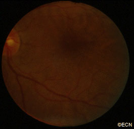
Let me explain Amblyopia: In order for an eye to achieve its best possible potential for vision, two things are necessary. First, proper images must be focused on the retina and second, the brain must receive those images. During the first 10 years of life, there is a process where images are collected and the brain learns to understand those images. If an eye is blocked by the tumor (or the eye lid), or if the eyes are not aligned, or if the eye is not able to focus images on the retina, the child’s eye-brain connection will not develop. That is, the child will not learn to see from that eye (a problem called amblyopia). Urgent treatment of periocular hemangioma of childhood can be necessary be to prevent amblyopia.

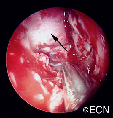
Periocular hemangioma of childhood has been treated with surgery, laser-surgery, radiation, and drugs (intralesional steroids and systemic beta-blockers). When possible, treatment of periocular hemangiomas of childhood involves injections of steroid into the tumor. In comparison to taking the medicine by mouth (PO) or by vein (IV), this approach has the advantage of putting the medicine right into the tumor. An acute effect, but does carry risk of tumor and orbital hemorrhage.

References

  1.  Haik B, Karcioglu Z, Gordon RA, Pechous BP. Capillary hemangioma (infantile periocular hemangioma). Survey of Ophthalmology 1994;38:399-426.
  2.  Kushner BJ. Hemangiomas. Archives of Ophthalmology 2001;118;835-836.

Related Links


Orbital Rhabdomyosarcoma

By Paul T. Finger, MD

Description

orbital_rhabdomyosarcoma
An Orbital Rhabdomyosarcoma

Rhabdomyosarcoma is the most common primary malignancy of the orbit in children. It can also occur in adults, though the average age of patients affected by rhabdomyosarcoma is 7 – 8 years.

Symptoms

Most parents first notice a droopy eyelid (called ptosis), and that the eye is more prominent (called proptosis), or that their child has a tumor under the conjunctival membrane that covers the eye (globe). Rhabdomyosarcoma is usually found in the superonasal orbit (that is under the upper lid near the nose).

orbital_rhabdomyoscaroma_ct
CT of Orbital Rhabdomyosarcoma

Diagnosis

Computed axial tomography (CT-scan) and magnetic resonance imaging (MRI) typically show a mass adjacent to or attached to one of the ocular or orbital muscles. CT is particularly helpful because it offers the best evidence if the orbital bones have been invaded by the rhabdomyosarcoma tumor.

Treatments

Rhabdomyosarcoma can grow rapidly and if the tumor grows into the brain or spreads to the lung, survival is poor. Prompt biopsy of a rhabdomyosarcoma followed by a combination of chemotherapy and irradiation offers the best chance of survival. In fact, recent reports suggest that current treatments offer greater than 90% survival from rhabdomyosarcoma.

Patients will develop problems typically seen after chemotherapy and irradiation of the eye, but if there is no recurrence after 3 years, it is likely that the rhabdomyosarcoma has been controlled.

orbital_rhabdomyosarcoma_histology
Histology of an Orbital Rhabdomyosarcoma

Related Links


Sclerosing Orbital Pseudotumor

By Paul T. Finger, MD

Description

sclerosing_orbital_pseudotumor
By local growth sclerosing orbital pseudotumors can cause bulging of the eye (proptosis).

Sclerosing orbital pseudotumor is uncommon. Due to unknown reasons, these tumor behave differently than other types of pseudotumor of the orbit. They grow more slowly, cause less pain, and are characterized by scarring (hardening of the tumor tissue). 

Symptoms

Sclerosing orbital pseudotumor is not cancer. But, by local growth it can cause bulging of the eye (proptosis), double vision (diplopia) and loss of vision. Sclerosing orbital pseudotumor can (rarely) extend into the sinuses, brain, and other orbit.

Diagnosis

sclerosing_orbital_pseudotumor_fibrous
Histopathology reveals large amounts of fibrous tissue.

Sclerosing orbital pseudotumor is usually diagnosed by biospy (orbitotomy).

Once the diagnosis is confirmed by pathology, systemic testing to rule out specific infectious and inflammatory causes should be performed. For example, an ANCA blood test and a chest x-ray should be performed to rule out Wegener’s Granulomatosis. Many of these patients have a past medical history of sinusitis, sinus surgery, or inhalation drug abuse. Therefore, concurrent treatable sinus disease should be addressed.

Treatments

In this case, computed tomographic (CT) scanning demonstrates a mass in the nasal orbit. The arrow demonstrates the optic nerve on stretch. The eye wall (sclera) is indented and the eye pushed out (proptosis).

Sclerosing orbital pseudotumors are less responsive to steroid therapy. Most cases are treated with combinations of surgery, steroid therapy, radiation and chemotherapy depending upon the clinical picture and the patient’s response to treatment.

Related Links


Orbital Pseudotumor

By Paul T. Finger, MD

Description

Inflammations can affect the tissues around the eye (orbit and adnexa). Certain orbital inflammations can look like tumors and are therefore called orbital pseudotumor. Orbital pseudotumor can affect one or both eyes of relatively young patients (less than 50 years old). They are not cancer.

Symptoms

Orbital pseudotumor can be quite painful. In fact, pain is one of the most prominent characteristics of this disease. In addition to pain, an inflammatory mass (tumor) can make the patient’s eye protrude (proptosis) and restrict the movement of the eye. A biopsy (called an orbitotomy) is commonly performed to confirm the diagnosis of orbital pseudotumor and to obtain tissue for pathology examination.

Diagnosis

Orbital pseudotumor is typically characterized by the rapid development of pain, proptosis, and swelling around the eye and orbit in adults. Ultrasound and computed tomographic (CT) scanning typically shows a diffuse infiltration of the orbit, an inflammation of the eye wall (sclera), and/or T-sign (with the optic nerve). Orbital pseudotumor related orbital masses typically have poorly defined margins. Systemic testing (blood and spinal fluid) may show signs of inflammation (e.g. increased sedimentation-rate) or atypical cells.

Patients with classic findings of orbital pseudotumor may be treated without a biopsy. A rapid complete response to steroid therapy helps confirm the diagnosis. Atypical cases of orbital pseudotumor usually undergo a diagnostic biopsy.

Specimens can be sent to test for infectious causes of orbital inflammation and certain systemic diseases. Typically eye cancer specialists will obtain blood, skin and radiographic (e.g. x-ray, MRI) tests for a variety of diseases such as sarcoidosis, tuberculosis, and Wegener’s Granulomatosis (see table below). An orbital biopsy can be particularly helpful in diagnosing many of these disorders.

Treatments

Orbital pseudotumor will respond rapidly to high-dose steroid therapy. Unfortunately, when the steroids are stopped, the inflammation often returns. Eye cancer specialists must reduce the steroid medication very slowly in order to prevent recurrence (return) of the disease.

In certain cases, chemotherapy (e.g. methotrexate, cyclosporine) and low-dose radiation (e.g. 1500-2500 cGy EBRT) may be needed to control the inflammation related to orbital pseudotumor. Most patients do well with steroid therapy but they are always at risk for recurrent orbital pseudotumor.

Additional info

Other Common Causes of Orbital Swelling and Inflammation:

  • Thyroid Eye Disease
  • Sarcoidosis
  • Infectious Orbital Cellulitis
  • Orbital Myositis
  • Scleritis
  • Orbital Vasculitis
  • Sjogren’s Disease
  • Wegener’s Granulomatosis
  • Malignant Ocular Tumors

Related Links


Orbital Mucocele

By Paul T. Finger, MD

Description

aadenoid_cystic
These magnetic resonance imaging (MRI) studies demonstrate displacement of the optic nerve, a bright T1 tumor image, and a variably bright T2 tumor image. The tumor is noted to involve the orbit, the ethmoid and frontal sinuses.

Orbital mucocele can occur when sinus mucoceles cannot naturally drain through the nose. Instead, they grow and slowly invade adjacent orbital tissues.

Generally arising from the ethmoid or frontal sinuses, orbital mucoceles are mucous or fluid filled cysts which can displace the eye. Frontal sinus mucoceles can force the eye down, ethmoid tumors will push the eye out and maxillary lesions can elevate or push the eye in. Lastly, orbital mucoceles originating in the sphenoid sinus can compress the optic nerve resulting in loss of vision.

Symptoms

Patients with mucocele of the orbit, typically have a history of facial trauma or chronic sinus disease. They tend to be in their mid 40’s or older. They can have painless proptosis, or complain of headaches, double vision, or loss of vision.

Diagnosis

orbital_mucocele_axial
Computed axial tomography demonstrates displacement of the medial rectus muscle (MR), as well as erosion and obliteration of portions of the orbital roof (black arrows). Note that the orbital portion of the mucocele is partially encased in bone. This is characteristic of mucocele. The rectus muscles and optic nerve are labeled.

demonstrates displacement of the medial rectus muscle (MR), as well as erosion and obliteration of portions of the orbital roof (black arrows). Note that the orbital portion of the mucocele is partially encased in bone. This is characteristic of mucocele. The rectus muscles and optic nerve are labeled.

Though magnetic resonance imaging (MRI) can be consistent with a mucous or serous fluid-filled tumor, a drainage procedure is typically required and found to be diagnostic. The mucoid or serous fluid (which is found to make up the mucocele) should be sent for culture and sensitivity as well as cytologic examination. Mucoceles can be infected. In those cases, the choice of antibiotics can depend on cultures taken during surgery.

Treatments

orbial_mucocele_transnasal
Intraoperative transnasal video photography is presented to show the mucocele cavity (arrow) after ethmoidectomy (photograph courtesy of Steven Schaefer, MD)

The treament of mucocele of the orbit is surgical. It is best to have a combination of an ophthalmic and ENT surgeons. Treatment involves removal of as much of the cyst and its lining as possible. This usually requires an orbitotomy and sinusectomy. It is most important to re-establish or create a new drainage pathway for the mucous to exit the nose.

Additional info

Case Example: A 63 year old male was referred to The New York Eye Cancer Center with a 6 month history of progressive painless proptosis of the right eye.

Despite this large orbital tumor with optic nerve displacement, the patient was 20/20 OU, he had no visual field defect, and no signs of optic neuropathy. A complete medical survey was initiated and the patient was cleared for surgery. A combination of anterior orbitotomy and transnasal ethmoidectomy were performed to evacuate the mucous and allow for future drainage.

Related links


Lymphangioma of the Orbit

By Paul T. Finger, MD
Computed Tomography (CT) shows a large lymphangioma (arrow), pushing the eye out of the orbit.

Description

Lymphangioma is rare (less than 7% of childhood orbital tumors). Patients can present with acute proptosis (bulging eye) after minor head trauma, as a gradual proptosis, or after an upper respiratory infection.

Symptoms

Lymphangioma tends to start in the superior and nasal orbital quadrants. More than 50% affect anterior (conjunctival and adnexal) structures. Typically, the lymphangioma bleeds into itself causing cysts of blood (called chocolate-cysts) within the tumor. If the cyst forms behind the eye, it pushes the eye forward. If the tumor forms in the eyelid or structures around the eye “adnexa”, blood filled lymphatic channels called “lymphangiectasias” can be seen beneath the conjunctiva

Diagnosis

A lymphangiectasia is seen beneath the conjunctival surface.

Lymphangioma is usually diagnosed by an eye cancer specialist. A careful history may reveal sudden painful proptosis (bulging of the eye), facial trauma or that the tumor or proptosis started right after a upper respiratory infection.

Physical examination may reveal bluish discoloration of or blood vessels within the eyelid skin. Should the vessels extend under the conjunctiva, they are called lymphangiectasias.

Severe cases can be associated with corneal exposure, ulceration and optic nerve damage.

Treatments

Though lymphangioma patients can present with a history of sudden proptosis (due to bleeding within the tumor), orbital lymphangiomas are typically slow growing. Therefore, most lymphangiomas are followed by observation for growth-related damage as documented by (clinical and radiographic studies) prior to intervention.

Thus, treatment of lymphangioma is indicated when associated with growth, optic nerve compression, corneal exposure problems (keratitis sicca), glaucoma or vision loss.

When treatment of lymphangioma is considered, the goal is rarely complete removal. This is because the edges of most orbital lymphangiomas are poorly defined. When necessary, patients undergo debulking surgeries with a goal of relieving acute optic nerve compression or corneal exposure. In rare cases, orbital lymphangioma patients may require orbital radiation therapy or exenteration for relief of pain.

Related Links


Cavernous Hemangioma of the Orbit

By Paul T. Finger, MD

Description

Computed tomography (CT) – Coronal Section demonstrates displacement of the optic nerve (medially) by the orbital hemangioma.

Hemangioma is a benign tumor that is found to grow within the orbit. Most commonly located behind the eye globe, it can push the eye forward causing eye-bulging doctors call proptosis.

Symptoms

Cavernous hemangioma of the orbit is most commonly seen in middle-aged women. Most are found within the muscle cone, but can be found anywhere in the orbit.

These orbital tumors can indent the back of the eye causing choroidal folds, or push on the optic nerve causing damage (atrophy).

Rarely, the tumor can push the eye so far that the cornea cannot be covered by the eye lids. In these cases, corneal exposure problems (keratitis, superficial punctate keratopathy, ulceration, even perforation) can occur.

Diagnosis

Cavernous hemangioma of the orbit is usually a slow-growing tumor. If the tumor has not damaged the eye, cavernous hemangioma can be observed for growth prior to considering intervention. Should tumor growth occur, it will be measured by eye examinations including (but not limited to) visual acuity, color vision assessment, Hertel exophthalmometry (a measure for proptosis), as well as an evaluation for double vision (strabismus), corneal exposure, retinal damage, vascular damage, and optic neuropathy.

Treatment

Treatment of orbital hemangioma is indicated when there is evidence of growth, optic nerve compression, and corneal exposure (with secondary keratitis sicca), or evidence of vision loss.

The goal of orbitotomy for choroidal hemangioma should be complete removal of the tumor. This usually involves careful dissection of the tumor to protect the tumor’s capsule (as possible). Connecting vascular feeder vessels should be identified and cauterized. A lateral orbitotomy can be required to keep the large tumor intact.

A fundus photograph demonstrates choroidal folds induced by the choroidal hemangioma indenting the posterior eye-wall.
Histopathologic sections with elastin-stain also demonstrate the large loosely distributed vascular channels associated with orbital hemangioma. The tumor’s capsule is noted on the right.

Adenoid Cystic Carcinoma of the Lacrimal Gland

By Paul T. Finger, MD

Description

Computed Radiographic Tomography (CT) demonstrates and Adenoid Cystic Carcinoma of the Lacrimal Gland with Orbital Extension (arrow)
Adenoid cystic carcinoma is a type of cancer that affects glandular structures. Around the eye there is a lacrimal (lac-kree-mall) gland that makes tears. Orbital adenoid cystic carcinoma usually occurs in patients 20-50 years old.

Symptoms

When an adenoid cystic carcinoma of the lacrimal gland grows, it typically pushes the eye down, towards the nose and forward. It can cause bulging of the eye (called proptosis). Another characteristic of adenoid cystic carcinoma is that it also invades local nerves causing pain. Therefore, pain and bulging of the eye are the most common symptoms of adenoid cystic carcinoma of the lacrimal gland.

Diagnosis

A complete eye examination with a clinical history and ophthalmic examination are crucial to the diagnosis of adenoid cystic carcinoma of the lacrimal gland and orbit.

CAT scans, MRI’s and ultrasounds are also helpful in determining the diagnosis. When the eye cancer specialist sees a well-defined tumor in the superior-temporal (upper – outer) part of the orbit, that may have eroded into adjacent bone and/or extend into the orbital apex, he or she should suspect the tumor might be an adenoid cystic carcinoma.

Other tumors to consider include: benign mixed tumor, adenocarcinoma, dacryoadenitis, or other processes affecting the lacrimal gland. Often times a biopsy is need to confirm the clinical diagnosis.

Treatments

When possible, an adenoid cystic carcinoma should be totally removed. This usually requires a surgery called a lateral orbitotomy. Unfortunately, total removal is often impossible due to the tumor’s size, shape, and presence of invasion. Should the adenoid cystic carcinoma be found to have a capsule, and should your doctor be able to remove it within its “capsule,” total excisional surgery offers the best prognosis.

However, in many cases the adenoid cystic carcinoma extends beyond the capsule making removal of the entire adenoid cystic carcinoma nearly impossible. It may require removal of the orbital contents, bones and adjacent structures. Due to the poor (local control) results from this type of extensive surgery; combinations of surgical removal, regional chemotherapy and local radiation therapy may be offered as an alternative.

Additional info


About Orbital Tumors: General Information

By Paul T. Finger, MD

Description

Tumors and inflammations can occur behind the eye. They often push the eye forward causing a bulging of the eye called proptosis.

Tumors and inflammations can occur behind and around the eye. They can push the eye forward causing a bulging of the eye called proptosis (image). Alternatively, if the bulging does not allow the eye lids to close, orbital tumor proptosis can cause corneal breakdown. The most common causes of proptosis are thyroid eye disease and lymphoid tumors (lymphoma and atypical lymphoid hyperplasia).

Other tumors include vascular tumors (e.g. hemangiomas, lymphangioma, hemangiopericytoma), lacrimal gland tumors (e.g. dacryoadenitis, benign mixed tumor, sarcoidosis and adenoid cystic carcinoma), and growths that extend from the sinuses into the orbit (e.g. squamous carcinoma, mucocele). Metastatic cancer  can come from other parts of the body to form an orbital tumor. Lastly, an orbit tumor can also be caused by inflammation (e.g. pseudotumor, sarcoidosis) or infection (abscess).

Symptoms

Most patients with orbital tumors notice a bulging of the eyeball or double vision (diplopia). Infections, inflammations and certain orbital cancers can cause pain. Less commonly, orbital tumors are accidentally discovered when patients have a CT or MRI of the head, sinuses and orbit.

Diagnosis

Though CT, MRI’s and ultrasound can help in determining the probable “clinical” diagnosis, most orbital tumors are diagnosed by a surgical biopsy called an orbitotomy (anterior or lateral). During biopsy a specimen is sent to an ophthalmic pathologist who helps determine the exact diagnosis. Dr. Finger has developed a small incision method of orbital biopsy called “FACT.”

Treatments

When possible, orbital tumors are totally removed. If they cannot be removed or if removal will cause too much damage to other important structures around the eye, a piece of tumor may be removed, sent for evaluation by a pathologist and the patient is treated with radiation and/or chemotherapy. Occasionally an orbital tumor is too big or involves the sinuses and requires more extensive surgery with bone-flaps.

If tumors cannot be removed during surgery, most orbital tumors can be treated with external beam radiation therapy. Certain rare orbital tumors require removal of the eye and orbital contents. However, in select cases alternative therapies (e.g. orbital radiotherapy and chemotherapy) can be used to treat residual tumor to spare vision and the eye.

Related links


Orbital Tumors


Patient Stories

"Very well treated by Dr. Finger. He explained everything I needed to know about my issue with detail and attention, putting me at ease and giving me confidence to handle this problem for the rest of my life.”
N.N.

Read More

REQUEST AN APPOINTMENT

Go to Appointment Form

CONNECT AND SHARE