Juxtapapillary Angioma Treatment Case

By Maria Saornil, MD

Color Photographs: Juxtapapillary Capillary Hemangioma

Dr. Soarnil would appreciate suggestions as to treatment options to optimize visual outcome without risk to this 42 year old woman with a juxtapapillary hemangioma.

“This is a case of juxtapapillary capillary hemangioma in a patient with V-H-L Disease, a woman 42 yo. She came to my clinic in February 2007, presenting a VA 20/20 in both eyes, no capillary hemangiomas in the RE and a juxtapapillary hemangioma in the left eye, asymptomatic. After 6 months, she started having visual symptoms and exudates (Top) and we performed TTT treatment. Exudates decreased, but an epiretinal membrane appeared. Hemangioma activity persisted, and a second TTT was performed December 2007, and ERM symptoms worsened and the hemangioma persisted.

View the fluorescein angiogram taken after TTT – treatment

Flourescein Angiography of the juxtapapillary hemangioma

What do you think is the next step? Intravitreal injection of anti- VEGF? Vitreoretinal surgery with endophotocoagulation ? Brachytherapy? Any combination? I will appreciate very much your suggestions.

Best regards,
Maria Saornil

Related Links


Orbital Mucocele

By Paul T. Finger, MD

Description

aadenoid_cystic
These magnetic resonance imaging (MRI) studies demonstrate displacement of the optic nerve, a bright T1 tumor image, and a variably bright T2 tumor image. The tumor is noted to involve the orbit, the ethmoid and frontal sinuses.

Orbital mucocele can occur when sinus mucoceles cannot naturally drain through the nose. Instead, they grow and slowly invade adjacent orbital tissues.

Generally arising from the ethmoid or frontal sinuses, orbital mucoceles are mucous or fluid filled cysts which can displace the eye. Frontal sinus mucoceles can force the eye down, ethmoid tumors will push the eye out and maxillary lesions can elevate or push the eye in. Lastly, orbital mucoceles originating in the sphenoid sinus can compress the optic nerve resulting in loss of vision.

Symptoms

Patients with mucocele of the orbit, typically have a history of facial trauma or chronic sinus disease. They tend to be in their mid 40’s or older. They can have painless proptosis, or complain of headaches, double vision, or loss of vision.

Diagnosis

orbital_mucocele_axial
Computed axial tomography demonstrates displacement of the medial rectus muscle (MR), as well as erosion and obliteration of portions of the orbital roof (black arrows). Note that the orbital portion of the mucocele is partially encased in bone. This is characteristic of mucocele. The rectus muscles and optic nerve are labeled.

demonstrates displacement of the medial rectus muscle (MR), as well as erosion and obliteration of portions of the orbital roof (black arrows). Note that the orbital portion of the mucocele is partially encased in bone. This is characteristic of mucocele. The rectus muscles and optic nerve are labeled.

Though magnetic resonance imaging (MRI) can be consistent with a mucous or serous fluid-filled tumor, a drainage procedure is typically required and found to be diagnostic. The mucoid or serous fluid (which is found to make up the mucocele) should be sent for culture and sensitivity as well as cytologic examination. Mucoceles can be infected. In those cases, the choice of antibiotics can depend on cultures taken during surgery.

Treatments

orbial_mucocele_transnasal
Intraoperative transnasal video photography is presented to show the mucocele cavity (arrow) after ethmoidectomy (photograph courtesy of Steven Schaefer, MD)

The treament of mucocele of the orbit is surgical. It is best to have a combination of an ophthalmic and ENT surgeons. Treatment involves removal of as much of the cyst and its lining as possible. This usually requires an orbitotomy and sinusectomy. It is most important to re-establish or create a new drainage pathway for the mucous to exit the nose.

Additional info

Case Example: A 63 year old male was referred to The New York Eye Cancer Center with a 6 month history of progressive painless proptosis of the right eye.

Despite this large orbital tumor with optic nerve displacement, the patient was 20/20 OU, he had no visual field defect, and no signs of optic neuropathy. A complete medical survey was initiated and the patient was cleared for surgery. A combination of anterior orbitotomy and transnasal ethmoidectomy were performed to evacuate the mucous and allow for future drainage.

Related links


Finger’s Slotted Eye Plaque for Treatment of Tumors Touching or Surrounding (Circumpapillary) the Optic Nerve

Finger's Slotted Plaque: Example 1
Finger’s Slotted Plaque: Example 1

Dr. Finger invented “Slotted Eye Plaques” for treatment of juxtapapillary and circumpapillary intraocular tumors. Standard COMS-type gold eye-plaques were altered such that an 8-mm-wide (variable length) slot was created to accommodate the orbital portion of the optic nerve. As the nerve entered the slot, the plaque’s posterior margin extended beyond the optic disk as to cover tumor (on the opposite side). Radioactive seeds around the slot, surround the tumor and its posterior tumor margins.

Dr. Finger says, “due to the physical limitations of notched plaques, there was no plaque device that could extend to cover and treat the posterior portion of a tumor that came within 1.5 mm, touched or surrounded the optic nerve.” This is why Dr. Finger invented slotted eye plaques. Intraoperative ultrasound imaging is used to confirm proper plaque placement. Preoperative radiation dosimetry showed that all tumour-tissue received a minimum of 85Gy (despite the gap created by the slot). To date, there has been only one tumor regrowth which is much better than previous plaque treatments for these tumors. No new complications that might prevent the use of slotted plaque radiation therapy have been noted.

Graphic Animation of a Slotted Plaque in Position over a Circumpapillary Tumor

Dr. Finger says, “slotted plaques represent an innovation that improves radiation coverage for tumors that touch or surround the optic nerve. For the first time, this technique allows plaque radiation therapy to include the entire juxtapapillary and circumpapillary tumor within the treated zone. It “normalizes” plaque treatment of these tumors. He adds, this technique addresses the problems some centers were having with local control of tumors in this location. Certainly, it will allow more patients to keep their eyes and their vision.”

For more complete information read these manuscripts published in the British Journal of Ophthalmology and Ophthalmology in 2012 or contact Dr. Finger.

Related Links


Optic Nerve Sheath Meningioma

By Paul T. Finger, MD

Graphic of how an eye can look with a posterior orbital tumor.

Orbital and optic nerve meningioma can extend from the brain into the orbit (behind the eye) and push the eye forward causing a bulging of the eye called proptosis. Though rare, when they occur, they are a significant cause of vision loss.

Symptoms

Patient with orbital meningioma typically have proptosis (bulging eye). Optic nerve compression can cause optociliary shunt vessels to form, as well as loss of vision. Depending on the location, size and degree of optic nerve involvement; patients can develop monocular and/or junctional defect is the patients field of vision.

Diagnosis

Opto-ciliary shunt vessels resulting from optic nerve compression

Patients usually present in their 40s and may have neurofibromatosis type 2. In making this diagnosis, one should look for the triad of vision loss, optic atrophy and abnormal vessels on the optic nerve. The nerve head can appear raised. Enlarged blood vessels are called “optociliary shunt vessels” and indicated that the meningioma has disrupted the natural circulation through the optic nerve to the retina and choroid. Angiography of the optic nerve head will clearly demonstrate the abnormal blood vessels. Magnetic resonance imaging (MRI), ultrasonography and computed tomographic (CT) imaging have been used to evaluate the orbital tumor and measure the optic nerve sheath diameter. CT is particularly helpful for imaging calcium within the tumor.


Computed tomography (CT) shows that the eye is pushed forward by this optic nerve sheath meningioma. Notice that the tumor is relatively bright (radio-dense). Imaging of the brain can determine if the meningioma extends into the brain.

In the images above, note that computed tomography of this optic nerve sheath meningioma. C demonstrates the calcific density of the left optic nerve. An x-ray film shows the linear density (seen on the right side of the film crossing the inferior orbital rim) which corresponds to the optic nerve sheath meningioma seen above.

Coronal "C-scan" ultrasound can be used to compare optic nerve diameters in a case of optic nerve sheath meningioma
Coronal “C-scan” ultrasound can be used to compare optic nerve diameters in a case of optic nerve sheath meningioma

Treatment Plan

Orbital meningioma is typically a slow-growing tumor. Once diagnosed, meningioma can be observed for growth prior to considering intervention. Treatment is indicated when there is a risk of spread to the central nervous system (in primary optic nerve sheath meningioma), documented progressive vision loss, or for rapid growth.

Computed tomography of the optic nerve sheath meningioma. Note the almost calcific density of the left optic nerve. An x-ray film shows the linear density (seen on the right side of the film crossing the inferior orbital rim) which corresponds to the optic nerve sheath meningioma seen above.

Though microsurgical resections have been tried (in an effort to spare the optic nerve), most eventually fail. The goal of local resection should be complete removal of the meningioma. This usually involves removal of the involved optic nerve. If complete surgical removal is not possible or in special circumstances, radiation therapy is commonly employed.

Biopsy:

Indications for biopsy include: atypical tumors, aggressive disease, acute vision loss and when a pathology diagnosis is requested. Certain inflammatory tumors can have a similar appearance to orbital meningiomas. However, biopsy carries risk for vision loss.

Treatment:

Orbital meningioma is typically a slow-growing tumor. One must consider patient age, rate of tumor growth and risk for loss of vision. That said, once diagnosed slow or non-growing meningiomas can be observed for further growth or stabilization prior to considering intervention. In general, treatment is indicated when there is a risk of spread to the central nervous system (in primary optic nerve sheath meningioma), documented progressive vision loss, or for rapid growth.

Treatment alternatives are tailored to the clinical situation. For example, if vision is lost, the tumor is growing toward the central nervous system and is resectable; it is removed. If complete surgical removal is not possible or in special circumstances,  microsurgical resection or external beam radiation therapy can be considered. These decisions are complex and best made with your neuro-ophthalmologist, neurologist and orbital surgeon.

Related Links


Lymphangioma of the Orbit

By Paul T. Finger, MD
Computed Tomography (CT) shows a large lymphangioma (arrow), pushing the eye out of the orbit.

Description

Lymphangioma is rare (less than 7% of childhood orbital tumors). Patients can present with acute proptosis (bulging eye) after minor head trauma, as a gradual proptosis, or after an upper respiratory infection.

Symptoms

Lymphangioma tends to start in the superior and nasal orbital quadrants. More than 50% affect anterior (conjunctival and adnexal) structures. Typically, the lymphangioma bleeds into itself causing cysts of blood (called chocolate-cysts) within the tumor. If the cyst forms behind the eye, it pushes the eye forward. If the tumor forms in the eyelid or structures around the eye “adnexa”, blood filled lymphatic channels called “lymphangiectasias” can be seen beneath the conjunctiva

Diagnosis

A lymphangiectasia is seen beneath the conjunctival surface.

Lymphangioma is usually diagnosed by an eye cancer specialist. A careful history may reveal sudden painful proptosis (bulging of the eye), facial trauma or that the tumor or proptosis started right after a upper respiratory infection.

Physical examination may reveal bluish discoloration of or blood vessels within the eyelid skin. Should the vessels extend under the conjunctiva, they are called lymphangiectasias.

Severe cases can be associated with corneal exposure, ulceration and optic nerve damage.

Treatments

Though lymphangioma patients can present with a history of sudden proptosis (due to bleeding within the tumor), orbital lymphangiomas are typically slow growing. Therefore, most lymphangiomas are followed by observation for growth-related damage as documented by (clinical and radiographic studies) prior to intervention.

Thus, treatment of lymphangioma is indicated when associated with growth, optic nerve compression, corneal exposure problems (keratitis sicca), glaucoma or vision loss.

When treatment of lymphangioma is considered, the goal is rarely complete removal. This is because the edges of most orbital lymphangiomas are poorly defined. When necessary, patients undergo debulking surgeries with a goal of relieving acute optic nerve compression or corneal exposure. In rare cases, orbital lymphangioma patients may require orbital radiation therapy or exenteration for relief of pain.

Related Links


Cavernous Hemangioma of the Orbit

By Paul T. Finger, MD

Description

Computed tomography (CT) – Coronal Section demonstrates displacement of the optic nerve (medially) by the orbital hemangioma.

Hemangioma is a benign tumor that is found to grow within the orbit. Most commonly located behind the eye globe, it can push the eye forward causing eye-bulging doctors call proptosis.

Symptoms

Cavernous hemangioma of the orbit is most commonly seen in middle-aged women. Most are found within the muscle cone, but can be found anywhere in the orbit.

These orbital tumors can indent the back of the eye causing choroidal folds, or push on the optic nerve causing damage (atrophy).

Rarely, the tumor can push the eye so far that the cornea cannot be covered by the eye lids. In these cases, corneal exposure problems (keratitis, superficial punctate keratopathy, ulceration, even perforation) can occur.

Diagnosis

Cavernous hemangioma of the orbit is usually a slow-growing tumor. If the tumor has not damaged the eye, cavernous hemangioma can be observed for growth prior to considering intervention. Should tumor growth occur, it will be measured by eye examinations including (but not limited to) visual acuity, color vision assessment, Hertel exophthalmometry (a measure for proptosis), as well as an evaluation for double vision (strabismus), corneal exposure, retinal damage, vascular damage, and optic neuropathy.

Treatment

Treatment of orbital hemangioma is indicated when there is evidence of growth, optic nerve compression, and corneal exposure (with secondary keratitis sicca), or evidence of vision loss.

The goal of orbitotomy for choroidal hemangioma should be complete removal of the tumor. This usually involves careful dissection of the tumor to protect the tumor’s capsule (as possible). Connecting vascular feeder vessels should be identified and cauterized. A lateral orbitotomy can be required to keep the large tumor intact.

A fundus photograph demonstrates choroidal folds induced by the choroidal hemangioma indenting the posterior eye-wall.
Histopathologic sections with elastin-stain also demonstrate the large loosely distributed vascular channels associated with orbital hemangioma. The tumor’s capsule is noted on the right.

Adenoid Cystic Carcinoma of the Lacrimal Gland

By Paul T. Finger, MD

Description

Computed Radiographic Tomography (CT) demonstrates and Adenoid Cystic Carcinoma of the Lacrimal Gland with Orbital Extension (arrow)
Adenoid cystic carcinoma is a type of cancer that affects glandular structures. Around the eye there is a lacrimal (lac-kree-mall) gland that makes tears. Orbital adenoid cystic carcinoma usually occurs in patients 20-50 years old.

Symptoms

When an adenoid cystic carcinoma of the lacrimal gland grows, it typically pushes the eye down, towards the nose and forward. It can cause bulging of the eye (called proptosis). Another characteristic of adenoid cystic carcinoma is that it also invades local nerves causing pain. Therefore, pain and bulging of the eye are the most common symptoms of adenoid cystic carcinoma of the lacrimal gland.

Diagnosis

A complete eye examination with a clinical history and ophthalmic examination are crucial to the diagnosis of adenoid cystic carcinoma of the lacrimal gland and orbit.

CAT scans, MRI’s and ultrasounds are also helpful in determining the diagnosis. When the eye cancer specialist sees a well-defined tumor in the superior-temporal (upper – outer) part of the orbit, that may have eroded into adjacent bone and/or extend into the orbital apex, he or she should suspect the tumor might be an adenoid cystic carcinoma.

Other tumors to consider include: benign mixed tumor, adenocarcinoma, dacryoadenitis, or other processes affecting the lacrimal gland. Often times a biopsy is need to confirm the clinical diagnosis.

Treatments

When possible, an adenoid cystic carcinoma should be totally removed. This usually requires a surgery called a lateral orbitotomy. Unfortunately, total removal is often impossible due to the tumor’s size, shape, and presence of invasion. Should the adenoid cystic carcinoma be found to have a capsule, and should your doctor be able to remove it within its “capsule,” total excisional surgery offers the best prognosis.

However, in many cases the adenoid cystic carcinoma extends beyond the capsule making removal of the entire adenoid cystic carcinoma nearly impossible. It may require removal of the orbital contents, bones and adjacent structures. Due to the poor (local control) results from this type of extensive surgery; combinations of surgical removal, regional chemotherapy and local radiation therapy may be offered as an alternative.

Additional info


About Orbital Tumors: General Information

By Paul T. Finger, MD

Description

Tumors and inflammations can occur behind the eye. They often push the eye forward causing a bulging of the eye called proptosis.

Tumors and inflammations can occur behind and around the eye. They can push the eye forward causing a bulging of the eye called proptosis (image). Alternatively, if the bulging does not allow the eye lids to close, orbital tumor proptosis can cause corneal breakdown. The most common causes of proptosis are thyroid eye disease and lymphoid tumors (lymphoma and atypical lymphoid hyperplasia).

Other tumors include vascular tumors (e.g. hemangiomas, lymphangioma, hemangiopericytoma), lacrimal gland tumors (e.g. dacryoadenitis, benign mixed tumor, sarcoidosis and adenoid cystic carcinoma), and growths that extend from the sinuses into the orbit (e.g. squamous carcinoma, mucocele). Metastatic cancer  can come from other parts of the body to form an orbital tumor. Lastly, an orbit tumor can also be caused by inflammation (e.g. pseudotumor, sarcoidosis) or infection (abscess).

Symptoms

Most patients with orbital tumors notice a bulging of the eyeball or double vision (diplopia). Infections, inflammations and certain orbital cancers can cause pain. Less commonly, orbital tumors are accidentally discovered when patients have a CT or MRI of the head, sinuses and orbit.

Diagnosis

Though CT, MRI’s and ultrasound can help in determining the probable “clinical” diagnosis, most orbital tumors are diagnosed by a surgical biopsy called an orbitotomy (anterior or lateral). During biopsy a specimen is sent to an ophthalmic pathologist who helps determine the exact diagnosis. Dr. Finger has developed a small incision method of orbital biopsy called “FACT.”

Treatments

When possible, orbital tumors are totally removed. If they cannot be removed or if removal will cause too much damage to other important structures around the eye, a piece of tumor may be removed, sent for evaluation by a pathologist and the patient is treated with radiation and/or chemotherapy. Occasionally an orbital tumor is too big or involves the sinuses and requires more extensive surgery with bone-flaps.

If tumors cannot be removed during surgery, most orbital tumors can be treated with external beam radiation therapy. Certain rare orbital tumors require removal of the eye and orbital contents. However, in select cases alternative therapies (e.g. orbital radiotherapy and chemotherapy) can be used to treat residual tumor to spare vision and the eye.

Related links


Systemic Chemotherapy

By Paul T. Finger, MD

Description

Chemotherapy commonly refers to the use of drugs to treat cancer of the eye, lids and orbit. Some of these drugs are also used to treat benign ocular tumors caused by inflammation.

Though this review tends to focus on the side-effects of chemotherapy, it is important to note that most patients are able to tolerate treatment, are helped by chemotherapy, and go on to enjoy their lives.

Clearly the use of chemotherapeutic drugs have cured patients and extended lives. Chemotherapy also offers a method to control inflammatory diseases and provide an alternative or adjunct to surgery and radiation.

How Chemotherapy Drugs Are Given

Systemic chemotherapy drugs can be infused into a patient’s vein (intravenous – IV) or by mouth (per oral – PO). These methods can expose the entire body to significant doses of chemotherapy.

In ophthalmology, we also give chemotherapy topically (ointment or drops) onto the eye. This places the drug on top of the tumor or affected area and limits the possibility of systemic (whole body) side effects. In ophthalmology, certain drugs are also given by periocular injection. The physician’s choice of delivery method depends on the type of chemotherapy drug and the reason why it is given.

Why Chemotherapy Drugs Are Given

Chemotherapy drugs are typically prescribed to attack or destroy a patient’s tumor or cancer. They can also be used to change a patient’s immune system. They can be used alone or in combination with radiation and/or surgery. In general, systemic chemotherapy is given when a patient needs a systemic (whole body) therapy, or when the risks of chemotherapy are less than those associated with radiation or surgery.

Side Effects & Complications

All drugs can have side-effects (unwanted effects). It is important to ask your doctor or nurse about the potential risks (side-effects) and benefits associated with your chemotherapy. This is important because each drug can have slightly different types of side-effects.

There are common side-effects associated with many chemotherapy drugs. Since these reactions may or may not be associated with your chemotherapy, you may want to ask if they will apply to your case.

A partial list of side effects includes: allergies, bone marrow depression, hair loss, mouth and throat problems, nausea and vomiting, sexual and reproductive problems, and skin changes.

Allergic Reactions

Allergic reactions can occur after taking almost any drug (including chemotherapy). They can occur immediately or can be delayed. If severe, the drug will be discontinued. If mild, allergy treatments – e.g. suppression agents (typically more drugs) can be used to proceed with chemotherapy.

Common allergic symptoms include:

  • Rashes on the skin
  • Swelling
  • Shortness of breath
  • Rapid heart beat

Rare allergic symptoms include:

  • Decreased blood pressure
  • Shock
  • Kidney failure

Since most chemotherapy is given in a clinic or hospital, patients are typically watched for reactions after drugs are administered. In these controlled environments, prompt treatment is administered and allergic reactions are treated.

When at home after treatment, patients can notice late allergic reactions. If you notice skin rashes, weakness while standing or sitting, progressive swelling or any unusual changes you should contact your physician immediately.

Bone Marrow Suppression

Bone marrow depression (suppression) is a side effect caused by certain chemotherapy drugs. When it occurs, it typically happens 7-10 days after the chemotherapy is started and usually recovers after 3 weeks. There are biotherapy medications that can speed bone marrow recovery.

Blood cells and other blood components are made in the bone marrow. Therefore, bone marrow depression results in decreased numbers of white blood cells, red blood cells, and platelets.

When the body loses white blood cells (leukopenia), it is a greater risk for infection.
When the body loses red blood cells (anemia), it can be associated with fatigue.
When the body loses platelets (thrombocytopenia), the blood has trouble clotting and the patient is at greater risk for bruising and bleeding.

Symptoms suggestive of bone marrow depression?

  • Fever or chills
  • Fatigue
  • Bruising or Bleeding

If you have fever, chills, fatigue, bruising or bleeding call your doctor immediately. In most cases your physician can give you medicines to treat bone marrow depression. These drugs can treat infections and compensate for low levels of white blood cells, red blood cells, and platelets.

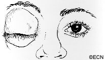
Hair Loss – Alopecia

Hair loss (alopecia) is a common side effect of chemotherapy, but not all chemotherapy drugs cause hair loss.

Not all hair loss is total. It can affect the head or all parts of the body. It can be gradual. Typically hair loss does not happen immediately. It starts several weeks after the first chemotherapy, then hair can thin or come out in clumps. Some people also experience scalp sensitivity.

It makes sense to use a mild shampoo, a soft hair brush, and low heat when drying your hair. Avoid dyes, perms and hair relaxers. In general, a short hair cut looks fuller. If you don’t cover your head, you may need to use sun screen on your scalp.

It is normal to feel angry or depressed when you lose your hair. You should speak with your doctor or nurse about your feelings. There may be a support group or mental health professional who can help you. But remember, hair loss is usually a temporary side-effect. The hair will grow back.

Hats and Wigs

Most sources say you should shop for a wig before you lose most of your hair. That way you can match your usual style and hair color. Consider getting a wig with tape-tab materials called “stickies.” This will allow you to comb and style the wig without worrying about sliding. Most sources also suggest you buy two wigs so you can have one on while the other is being cleaned.

Check to see if your insurance company will cover its cost. If not, it is a tax-deductible expense (keep your receipt). Have the establishment write, “hair prosthesis as prescribed by doctor” on the receipt.

When buying a hat, make sure it will block the sun from hitting your scalp. Women also prefer to wear hats or turbans when at home. Don’t forget your sunglasses.

Mouth and Throat Problems

Chemotherapy can cause sores, red areas or white patches to form on the mouth and throat. This is particularly true for systemic methotrexate and 5-fluorouracil.

Since mouth and throat care are known to improve, stabilize or prevent progression of mouth sores, patients should call their doctor if mouth sores or soreness occurs.

Typical Mouth Care Instructions May Include:

  • Regular, gentle cleaning of your teeth or dentures.
  • Also, rinse your mouth with a mixture:
    • 1/2 teaspoon of salt in 8 ounces of water or
    • 1/2 teaspoon of sodium bicarbonate (baking soda) in 8 ounces of water.
    • This should be done after every meal and at bedtime.
  • Avoid alcohol containing mouthwash.
  • Avoid acidic or spicy foods.
  • Avoid drinking alcohol and smoking.

Your doctor may have different methods of mouth care. So contact your doctor if you develop mouth sores, white patches, mouth pain or difficulty swallowing.

Nausea and Vomiting

Nausea and vomiting are the body’s defenses against eating too much and its mechanism to get rid of toxic food and poisons. During vomiting, the muscles that normally push food down the gastrointestinal tract, reverse direction and send back the partially digested material.

Nausea and vomiting may occur within the first hour of chemotherapy and can last up to a week. Most of the time nausea goes away shortly after these drugs are given.

In severe cases, anti-nausea medications may be necessary.
That is because the vomiting patient may lose too much fluid and necessary minerals. When a patient is at risk of dehydration and blood chemical (electrolyte) imbalance, your doctor or nurse can prescribe medication to help you prevent or lessen nausea.

Other causes of nausea that cancer patients may be experience include stress, radiation of the brain, and anesthetics (associated with surgery).

Treatment of Nausea and Vomiting 

The primary treatment for nausea is anti-nausea drugs. Since they work best taken before chemotherapy, discuss this with your doctor before your next treatment (particularly if you were nauseated after your last treatment).

Other methods include: acupressure wrist bands which are available at most drug stores, fasting (do not eat) for a couple of hours before your treatment, try to keep your eyes open, and slowly deep-breathing through your nose.

If you are already nauseated from chemotherapy, do not eat a large meal or drink carbonated beverages. You may try to eat crackers, pretzels, or dry toast. Avoid foods with a lot of residue, salads, high fiber cereals or fatty and fried foods.

Sometimes, right after chemotherapy patients are only able to suck on ice chips, sip ice water, or mild herbal tea. Once patients are feeling better, they may benefit from chicken soup.

If nausea and vomiting persist, call your doctor. Some patients need to receive intravenous (IV) fluids and electrolytes (minerals).

Sex and Reproduction

Chemotherapy-related sexual and reproductive problems can occur in both men and women.

Since certain chemotherapy drugs may have harmful effects on an unborn child, genetic counseling should be made available to the chemotherapy patient in order to discuss the effects of drug therapy on current and future pregnancies.

Studies & Research

  1. Wilson MW, Czechonska G, Finger PT, Rausen A, Hooper ME, Haik BG. Chemotherapy for Eye Cancer Survey of Ophthalmology 45:416-444, 2001.
  2. Calabresi P, Chabner BA. Chemotherapy of Neoplastic Diseases: Introduction. In: Goodman and Gilman’s: The Pharmacological Basis of Therapeutics. Tenth Edition Hardman JG, Limbird LE, Gilman AG (eds.) McGraw-Hill, New York, 2001, Section IX, pp.1381-1388.
  3. Chabner BA, Ryan DP, Paz-Ares L, Garcia-Carbonero R, Calabresi P. Antineoplastic Agents. In: Goodman and Gilman’s: The Pharmacological Basis of Therapeutics. Tenth Edition Hardman JG, Limbird LE, Gilman AG (eds.) McGraw-Hill, New York, 2001, Chapter 52, pp.1389-1460.

Proton Beam Therapy: What Patients Should Know!

Proton Beam Radiotherapy

By Paul T. Finger, MD

Proton beam radiation therapy is a form of teletherapy, where the radiation is directed into the eye from a machine outside the eye.

Proton Beam Radiotherapy- Graphic demonstration

In order to aim the tube-shaped proton beam, surgical clips are sewn (surgically placed) on to the eye, around the tumor’s base. Then the patient returns for 3 to 5 daily outpatient treatments. During treatment, the eye must not move. Eye movement displaces the column of radiation away from the tumor, causing unnecessary radiation of normal ocular structures. Therefore, the eye must be open and closely monitored for movement while the radiation beam is traveling through the eye. If the patient closes their eye, it naturally rotates up “Bell’s Phenomenon” changing the position of both the eye and its tumor.

Eye cancer specialists commonly suggest protons or other external beam treatments when the tumor is touching (juxtapapillary) or surrounding (circumpapillary) the optic nerve. Since beam therapy can include the entire tumor (plus a tumor-free margin), beam therapy is likely to destroy the tumor. However such treatment places the optic nerve within the targeted radiation zone that also results in radiation-related optic nerve damage and loss of vision (in that eye).

Proton beam typically involves directing a column of radiation through the front of the eye (anterior segment, eye lids and/or orbit) in order to reach the intraocular tumor. Since the radiation typically enters the front of the eye, eyelash loss, eyelid excoriation, corneal neovascularization and ulceration, dry eye, neovascular glaucoma, and cataract are more common after treatment with proton beam radiation therapy (compared to low-energy plaque radiation therapy). Some of these effects may occur within weeks of treatment, others may take years to develop.

VS. Eye-Plaque Radiotherapy

Eye-Plaque Radiotherapy: Graphic demonstration
Eye-Plaque Radiotherapy: Graphic demonstration of plaque radiation as it moves through the eye in treatment of a posterior tumor.

In contrast, Plaque Therapy typically involves attaching a dish-shaped radiation source beneath the tumor and leaving it there for 5-7 days.

Compared with proton-beam, the front of the eye usually receives much less radiation with plaque radiation therapy, but parts of the back of the eye may receive more. This is why anterior “front of the eye” complications (eye lash loss, severe dry eye, neovascular radiation glaucoma) are unusual after low-energy palladium-103 or the older iodine-125 type ophthalmic plaque radiation therapy.

Tumors that touch the optic nerve are more difficult to treat with ophthalmic plaque radiation therapy. Over 12 years ago, Dr. Finger developed specialized “Finger’s slotted plaques” for treatment of tumors the touch or encircle the optic nerve (see below). Due to the advent of super-sized 24 mm (for extra-large tumors) and Finger’s slotted eye plaques (for tumors around the optic nerve), fewer than 6% of patients require enucleation (removal of the eye) as initial treatment for choroidal melanoma (at The New York Eye Cancer Center). Clearly, ophthalmic plaque radiation therapy is the most widely available and most commonly used eye and vision-sparing treatment for choroidal melanoma.

An Overview of Plaque Radiation Therapy in Treatment of Choroidal Melanoma (1,2)

*Note* These results were published largely prior to the discovery of intraocular anti-VEGF therapy for radiation retinopathy and optic neuropathy. To see Dr. Finger’s current results, go to his results page on this web site.

Plaque Therapy Chart: Published results after several forms of plaque radiation therapy.

Summary

Currently eye cancer specialty centers should be able to offer radiation as an eye and vision-sparing alternative for 95% patients with intraocular melanoma. The two main types of radiation are eye-plaque brachytherapy (iodine-125, ruthenium-106, palladium-103) and external beam (Proton-Beam, stereotactic radiosurgery). Radioactive plaques are more commonly used and available in more centers throughout the world.

Side effects/complications

An Overview of Plaque Radiation Therapy in Treatment of Choroidal Melanoma (1,2)

Related links

References

  1. Finger PT. Radiation Therapy for Choroidal Melanoma. Survey of Ophthalmology (Review Article) 42:215-32, 1997.
  2. Finger PT., Berson A, Ng T, Szechter A. Palladium-103 Plaque Radiotherapy for Choroidal Melanoma: An 11-year study. Int. J Radiation Oncology Biol. Phys. Vol 54, No. 5, pp. 1438-1445, 2002.

Patient Stories

"Very well treated by Dr. Finger. He explained everything I needed to know about my issue with detail and attention, putting me at ease and giving me confidence to handle this problem for the rest of my life.”
N.N.

Read More

REQUEST AN APPOINTMENT

Go to Appointment Form

CONNECT AND SHARE