Chronic Lymphocytic Leukemia

By Paul T. Finger, MD

Description

Leukemic Infiltrate of the Anterior Chamber - Hypopyon with Hyphema
Leukemic Infiltrate of the Anterior Chamber – Hypopyon with Hyphema in a 6-year old child

Leukemia is a systemic disease that can involve the eyes. It occurs in both children and adults. In that the eyes can be a relative sanctuary (a place to hide from systemic chemotherapy) for leukemic cells, intraocular recurrences have been reported after both total body irradiation and systemic chemotherapy. It is reasonable for all patients with leukemia to have periodic eye examinations.

Adults can also have leukemia and develop infiltration of the eyes. The case presented in this section (below) demonstrates one such case.

Symptoms

Depending on the location of the leukemic infiltrates, patients will have different symptoms. Most patients are found to have retinal tumors with hemorrhage, optic nerve swelling (papilledema) or cells in the anterior chamber of the eye (hypopyon/hyphema). Leukemic tumor called “chloroma” can also form around the eyes and in the orbit.

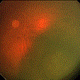
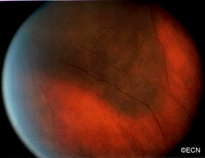
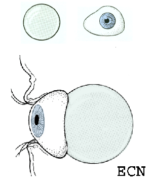
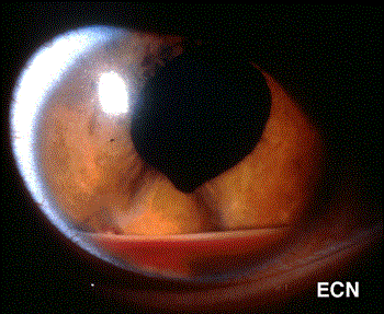
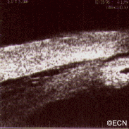
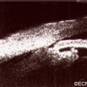
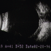
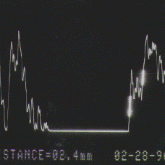
In this case, posterior uveal infiltration caused thickening of the choroid that moved the retina forward. This made the patient more far-sighted (hyperopic shift) due to a shortening of the distance between the cornea/lens complex and the retina.

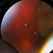
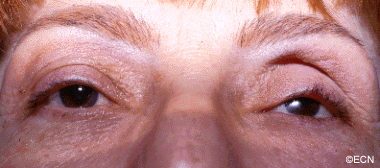
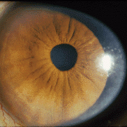
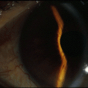
The front of the eye was also infiltrated by leukemic cells. Thickening of the iris, ciliary body and anterior uvea induced a secondary “narrow angle” glaucoma. The symptoms of glaucoma include seeing haloes around lights, headache (brow-ache) and eye pain.

Diagnosis

The clinical diagnosis of infiltrative ocular leukemia (e.g. typical findings and a history of systemic leukemia) is typically adequate for treatment of patients. When proof of recurrence is needed to proceed with systemic or local therapy, a biopsy may be performed for a cytologic or histopathologic diagnosis.

Treatments

The treatment of intraocular leukemia is (in part) dependent on the treatment of the patient’s systemic disease. For example, in the clinical situation where the eye findings are not posing an immediate threat to vision, your physician my wait for systemic therapies to resolve the ocular disease. In cases where all standard treatments have been unsuccessful in resolving the ocular leukemia; patients will be treated with low-dose external beam radiation therapy.

Related links


Squamous Carcinoma of the Eyelid

By Paul T. Finger, MD


Note the relatively flat surface, red (indurated – inflammed edges, and the white flakey material on the right side of its surface.

Description

Squamous carcinomas of the eyelid can locally invade the orbit and sinuses, but rarely metastasizes. It is the second most common malignant eyelid tumor, but is 10 times less common than basal cell carcinoma. This should not be confused with conjunctival squamous carcinoma, which is the most common conjunctival cancer and can spill over onto the eyelid skin.

Symptoms

Squamous eyelid carcinoma can have symptoms that range from the appearance of a hypervascular flat pale, reddish or flaky lesion on the eyelid skin to a thickened well-demarcated reddish, flat tumor surrounded by inflammation (with or without scaling from its surface).

Diagnosis

Squamous carcinoma of the eyelid should be photographed at baseline. These lesions can remain unchanged (for years), then invade into the dermis and grow. A simple wedge biopsy can be performed and sent for pathologic evaluation. Once the diagnosis of squamous carcinoma is biopsy proven, definitive treatment is needed.

Treatments

Like basal cell carcinomas, squamous cell cancers of the eyelid rarely metastasize. However, they can grow around the eye into the orbit, sinuses and brain. Therefore, early intervention with complete excision or destruction is warranted.

Surgical approaches depend on the doctors training. Eye care specialists will either perform a planned excision with frozen-section control (of the margins) or the Moh’s Technique. Both types of surgery are used to remove the entire tumor along with a safety zone of normal appearing tissue from the edges of surgical wound (margins). No comparative studies have definitively shown that one technique better than the other. Both surgeries require a cosmetic surgical repair to return eyelid function and cosmesis. When the orbit and sinuses are not involved, local excision is usually curative.

Extension into the orbit and sinuses typically requires more extensive surgery (exenteration, sinusectomy) with subsequent radiation therapy.

At the New York Eye Cancer Center, we have treated select patients with small squamous carcinomas of the eyelid with topical chemotherapy agents or freezing (cryotherapy) using “Finger-tip” applicators. Dr. Finger tries to avoid surgery when possible.

Related links


Sebaceous Carcinoma of the Eyelid

By Paul T. Finger, MD

Description

Sebaceous carcinoma arises from the glands within the eyelids, caruncle or eyebrow. They are more commonly found on the upper eyelid and in middle-aged patients.

Symptoms

Sebaceous cell carcinoma is suspected due to evidence of eyelash loss and the formation of a yellow-nodule. This tumor can also present as a persistent (months) non-responsive blepharitis or conjunctivitis. In these cases, a high index of suspicion for sebaceous cell carcinoma will lead to biopsy and the diagnosis. Once sebaceous carcinoma is suspected a biopsy is warranted. Before surgery, the pathologist should be advised of this possible diagnosis so the specimen can be processed appropriately

Sebaceous carcinomas are one of the rarest eye cancers and can look like a chalazion (stye) or conjunctivits. In that sebaceous carcinoma can mimick these relatively benign dieases, eye care specialists should be suspicious of this tumor in any patient with persistent conjunctivitis, blepharoconjunctivitis or chronic/recurrent chalazion. Therefore, any conjunctivitis or chalazion that is not getting better after 3 months of observation, should be biopsied.

Diagnosis

Sebaceous carcinoma of the eyelid typically presents as a yellow-nodule in the upper lid. The patient may have history of colon, breast, genitourinary or other skin malignancies (Muir-Torre Syndrome).* When a nodular tumor, it can cause eyelash loss, but is not painful (compared to styes). This tumor can also present as a hypervascular conjunctival tumor and tends to spread along the conjunctival surface. In these cases sebaceous carcinoma looks like conjunctivitis.

It is worth repeating that “any conjunctivitis or chalazion that is not getting better after 3 months of observation should be biopsied.” If cytopathology is available at your medical center, a scrape biopsy of the conjunctiva can be initially performed in the office setting. However, the gold standard for diagnosis of sebaceous carcinoma is histopathologic evaluation of a fresh tissue specimen.

Once the diagnosis is made, a metastatic survey is warranted. Sebaceous carcinoma can spread to regional lymph nodes (pre-auricular and cervical) as well as to lungs, brain, liver and bone. At The New York Eye Cancer Center, we obtain a total body PET/CT scan (from head to toes) for staging. Published series have reported that the occurrence of metastatic disease was dependent on the size and location of the primary tumor and occurred in up to 27% of patients. Local tumor invasion of the lymph nodes, orbit or metastatic sites were associated with a poor prognosis for survival.

Treatments

Biopsy usually requires a full thickness lid section sent for histopathologic evaluation prior to formalin fixation. Histopathologic evaluation reveals foamy cytoplasm (with hematoxylin and eosin) This specimen is found to be oil-red-o positive, consistent with sebaceous carcinoma. *note* Sebaceous carcinomas may occur several years before a gastrointestinal malignancy. This occurrence has been described as the autosomal dominant - Muir-Torre Syndrome.
Biopsy usually requires a full thickness lid section sent for histopathologic evaluation prior to formalin fixation. Histopathologic evaluation reveals foamy cytoplasm (with hematoxylin and eosin) This specimen is found to be oil-red-o positive, consistent with sebaceous carcinoma. *note* Sebaceous carcinomas may occur several years before a gastrointestinal malignancy. This occurrence has been described as the autosomal dominant – Muir-Torre Syndrome.

Treatment requires a resection of the malignancy. Frozen section control can be used to provide negative margins. Larger surgeries, cryodestruction and radiation may be required if resection is not possible. Exenteration (complete removal of the orbital contents is sometimes required for extensive or recurrent disease).

* Muir-Torre Syndrome: A rare, genetically autosomal dominant cancer syndrome. Patients with Muir-Torre can develop cancer of the colon, breast, genitourinary tract, skin and eye lesions, including keratoacanthomas and sebaceous carcinoma.

Related links


Malignant Melanoma of the Eyelid

By Paul T. Finger, MD

Description

Melanoma of the eyelid is a relatively rare tumor making up less than 1% of eyelid cancers. However, like other skin melanomas it can spread to other parts of the body. This tumor typically appears as a pigmented thickening (tumor) of the eyelid or extension of pigment from the conjunctiva. It is characterized by a patient history of or photographically documented growth.

Symptoms

Malignant melanoma of the eyelid may look like an eyelid nevus, but it more likely to be  variably pigmented, change color, bleed and/or grow. All pigmented eyelid tumors of the eyelid should be evaluated by an eye cancer specialist.

Diagnosis

All eyelid tumors should be photographed for comparison with future examinations. if possible, patients should keep a copy of their baseline photographs. Malignant melanoma of the eyelid is distinguished from an eyelid nevus in that it can be variably pigmented, change color, bleed and/or grow. If your doctor makes a clinical diagnosis of eyelid melanoma or change is noted, the tumor should be evaluated by an incisional (not superficial) biopsy. This is because the depth of the tumor is an important risk factor for spread to the rest of the body.  A simple wedge biopsy can be sent for pathologic evaluation to aid in the diagnosis. To evaluate the entire tumor, primary excisional biopsies need be performed. Once the diagnosis is made on pathology, systemic staging with total body, scalp to toes PET/CT imaging should be performed.

Treatments

When the pathology diagnosis confirms or discovers that the tumor is malignant melanoma of the eyelid (eyelid cancer), a medical work-up is ordered to stage the patient and thus rule out metastatic spread to other parts of the body. If there is no metastatic disease, the tumor can be surgically removed (including large margins of normal appearing tissue). At the time of surgery, some doctors will also remove regional lymph nodes near the tumor (in order to determine if the tumor has locally spread).

Ophthalmic plastic surgery techniques are usually required to repair the defect caused by tumor removal. The extent of surgery is governed by the ocular distribution of the tumor. That said, it can range from simple eyelid resection (wedge or Moh’s) to removal of the eyelid, debulking the orbit, to orbital exenteration. Radiation (external beam or brachytherapy implant) may be added if surgical margins are not possible.  Most all patients can be repaired using one of these techniques (see Treatment Types Section).

Related links


Benign Eyelid Tumors – Seborrheic Keratosis

By Paul T. Finger, MD

Description

Seborrheic keratosis is one of the most frequently noted benign eyelid tumors.

Symptoms

This is a superficial brown to black, cobble-stone surfaced tumor. Note its “stuck-on” appearance, its cobblestone surface, well-defined margins and lack of inflammation. Histopathology reveals a hyperkeratotic tumor consisting of a basaloid proliferation with keratin cysts.

Unlike squamous carcinomas of the eyelid, seborrheic keratosis does not typically cause inflammation around its edges nor does it have a smooth reddened scaly surface. If it does become inflamed it can be mistaken for a basal or squamous cell carcinoma.

Diagnosis

Typical appearing seborrheic keratosis can be photographed and followed for evidence of change prior to consideration of biopsy or removal. Suspicious seborrheic keratosis can be differentiated from other eyelid tumors by a simple biopsy.

Treatments

Seborrheic keratosis does not require treatment. Photography should be used to document its initial size for future reference and comparison. Suspicious lesions should be biopsied or removed to differentiate them from malignant eyelid tumors.

Note its “stuck-on” appearance, its cobblestone surface, well-defined margins and lack of inflammation. Histopathology reveals a hyperkeratotic tumor consisting of a basaloid proliferation with keratin cysts.

Related links


High Frequency Ultrasonography – A New View of Iris and Ciliary Body Tumors

By Paul T. Finger, MD

Case 1: Dynamic Scanning

Case 1: An Iris Melanoma after Dilation
Case 1: An Iris Melanoma before Dilation

Notice the low reflective mass in the iris. There is thinning of the iris pigment epithelium and a length of relatively normal appearing iris between the posterior margin of the tumor and the ciliary body. In this case, the tumor was documented to grow and cataract surgery was contemplated. In part, due to these dynamic high-frequency ultrasound findings, excision was planned prior to dilation for cataract surgery.

This case illustrates that high-frequency ultrasound allowed for unique views of the posterior aspect of the tumor, as well as an assessment of its invasion within the iris stroma.

Case 2: A Sigh of Relief

high_freq_case2_2
Case 2: High-frequency ultrasonography demonstrates a cystic “acoustically empty” tumor displacing the iris anteriorly, causing focal angle closure.
hight_freq_case2
Case 2: A smooth-surfaced tumor is seen posterior to the iris stroma and anterior to the anterior lens capsule.

The iris cysts are the most common iris tumor sent for high-frequency ultrasonographic evaluation. Its clinical presentation is similar to seeing a localized “bulge” in the iris. Ultrasonography clearly demonstrates the cystic nature of the tumor and allows for an assessment of the adjacent ciliary body (for tumor).

Cases 3 & 4: A Unique tool

high_freq_case3
Image 2
high_freq_case3_2
Image 1

This melanoma has grown through the iris pigment (image 1)  epithelium onto the anterior capsular surface. High frequency ultrasonography allows for an assessment of tumor penetration of the iris and ciliary body.

This small ciliary body melanoma (image 2) would likely have gone undetected prior to high frequency ultrasonography. Like other malignant tumors, early detection and treatment of ciliary body melanomas should improve survival.

 References

  1. Reminick LR, Finger PT, Ritch R, Weiss S, Ishikawa H. Ultrasound biomicroscopy in the diagnosis and management of anterior segment tumors. Journal of the American Optometric Association 69(5);575-582, 1998.
  2. Katz NR, Finger PT, McCormick SA, Tello C, Ritch R, Sirota M, Kranz O. Ultrasound biomicroscopy in the management of malignant melanoma of the iris. The Archives of Ophthalmology 1995;113:1462-1463.

Related Links


Diode-Light Transillumination

By Paul T. Finger, MD
diode-light_1Ophthalmic plaque brachytherapy has become the most common eye-sparing technique for the treatment of intraocular tumors. Eye plaques are constructed by affixing radioactive seeds within a gold, metallic, bowl-shaped carrier.

Then the radioactive eye plaque is sewn to the wall of the eye underneath the intraocular tumor. It is left in place for several days during which the tumor is sterilized by the radiation.

Small diode lights can be attached to ophthalmic plaques in order to improve their localization beneath their intraocular tumors and to allow photo-documentation of plaque position. With the lights turned on, we could see that the plaques were in position as to cover the intraocular tumor.

In most centers, radiation therapy has been able to destroy choroidal melanomas in over 92% of cases. Unfortunately, at least 8% of tumors have been found to regrow within 5 years of treatment. These failures of local control are thought to be due to radiation resistant tumors and to suboptimal placement of the eye-plaque (using standard transillumination techniques). This is evidenced by more failures occurring in treatment of small tumors near the optic nerve. These cases are difficult or impossible to visualize with standard transillumination or ultrasound techniques. Diode-light transillumination was invented to improve localization of eye plaques beneath these small posterior tumors.

References

  1. Finger PT, Iezzi R, Romero J, Rosen RB, Szechter A, Hegde H. Plaque-Mounted Diode-Light Transillumination For Localization around Intraocular Tumors. The Archives of Ophthalmology 1999:117:179-183.
  2. Finger PT, Iezzi R, Esteveo ML, Szechter A, Rosen RB, Berson A. Diode-Light Transillumination for Ophthalmic Plaque Localization around Juxtapapillary Choroidal Melanomas. The International Journal of Radiation Oncology, Biology, Physics 44:887-890, 1999.

Related Links


Hyperthermic Treatment of Intraocular Tumors

By Paul T. Finger, MD

Ten years after treatment the tumor has regressed to scar with scleral show.

Heat “Hyperthermia” has been used to make radiation more effective in the treatment of cutaneous and metastatic malignant melanoma. This is because relatively low levels of heat (42-45 Celsius) can inactivate enzymes used to repair radiation damage and higher temperatures can damage tumor blood vessels.It is believed that low level heating offers the potential to decrease the amount of radiation required to kill cancer cells resulting in fewer radiation complications.

The largest and longest followed series of patients treated with both eye-plaque radiation therapy and ocular hyperthermia was published in 1997. This phase-I clinical case series demonstrated comparable local control and improved visual acuities compared to other series of patients treated with radiation alone. For further information read, see links below.

Unfortunately, due to the small numbers of patients and commerical liability concerns/expense, hyperthermic treatment of choroidal melanoma was not found to be commercially viable. If there is commercial interest, please contact Dr. Finger.

References

An anterior choroidal melanoma prior to I-125 /microwave thermoradiotherapy.
  1. Finger PT. Microwave thermoradiotherapy for uveal melanoma: Results of a 10-year study. Ophthalmology 1997;104:1794-1803.
  2. Lagendijk JJ. A microwave heating technique for the hyperthermic treatment of tumours of the eye, especially retinoblastoma. Phys Med Biol 1982; 27:1313-1324.
  3. Petrovich Z, Pike M, Astrahan MA, Luxton G, Murphree AL, Liggett PE. Episcleral plaque thermoradiotherapy of posterior uveal melanomas. Am J Clin Oncol 1996;19:207-211.
  4. For a complete review of the methods of hyperthermic treatment for intraocular tumors read:  Finger PT. Thermoradiotherapy for Eye Tumors. In: Medical Radiology – Diagnostic Imaging and Radiation Oncology, Volume 2: Principles and Practice of Thermoradiotherapy and Thermochemotherapy. Seegenschmiedt MH, Fessenden P, Vernon CC, eds. Berlin: Springer-Verlag, 1993 175-185.

Related Links


Enucleation Implants: “Finger’s Expandable Orbital Implant”

By Paul T. Finger, MD

Innovation

Dr. Finger’s patented, expandable orbital implant will have the capacity to be enlarged as the surrounding orbital tissues atrophy. An enlarging prosthesis should help correct ptosis (drooping eyelid), enophthalmos (sunken eye) and motility (movement). Clinical trials will be needed to evaluate this effect on movement and cosmetic results for patients. The movie demonstrates the capacity of the implant to expand. In practice, the implant will be inflated and allowed to stay in its expanded state. Dr. Finger is interested in finding a company to produce this patented implant.

Advantages

  1. They cannot become infected.
  2. They are not made of nor contain calcium (that can affect subsequent imaging or radiation treatment). The implant sits in the orbit and behind the ocular prosthesis.

General Information About Enucleation

The expandable orbital implant has an anterior injection portal, rectus muscle placement grooves, and 4 independently expandable/deflatable quadrants.

Volume-Replacement After Enucleation

Enucleation (“removal of the eye”) is typically recommended for patients:

1. With a large intraocular eye cancer which cannot be destroyed or removed by other methods.
2. When alternative treatments are likely to leave the patient with poor or no useful vision.
3. With a blind and painful eye.
4. When a patient requests removal of the eye as treatment for their eye cancer.

Current Types of Implants

Drooping of the left upper eye lid (ptosis)

Implants are used to replace the volume lost by the enucleation procedure. 

When this type of implant is placed in the orbit, it is surrounded by the extraocular muscles and soft tissues. After it heals, the prosthesis (which looks much like a giant contact lens) is placed over the implanted ball. The ball-muscle complex functions much like the ball in a ball-in-socket joint. Therefore, when the implant moves, the overlying prosthesis also moves.

Non-integrated implants:

The implant sits in the orbit and behind the ocular prosthesis.
  1. The can be made of glass, PMMA, silicone (PMMA plastic is the most common)
  2. These are the least expensive
  3. They are inert, meaning they do not interact with orbital tissues
  4. They will not attach to orbital tissues, so they are more likely to move or become displaced within the orbit.

Integrated implants:

1. Pegging of the implant provides increased movement.
2. Larger implants can be used.
3. The muscles can be directly sewn to the implant.

For more information on enucleation you can go to a medical library, or click and order a copy of our comprehensive review of:

  • “Enucleation” by Moshfeghi DM, Moshfeghi AA, Finger PT. Survey of Ophthalmology 2000:44:277-301.
Hydroxyapetite Orbital Implants

Related Links


Dr. Finger Representing All Eye Cancer Specialists At The Commission On Cancer Spring Meeting 2016

Dr. Finger recently attended the 2016 Commission on Cancer Spring Meeting in Chicago. It was an opportunity to not only network with other cancer doctors from around the United States, but to plan and move forward a number of initiatives that will benefit the eye cancer care community and its patients.

COCSignFinger

Dr. Finger serves as a representative of the American Joint Committee on Cancer (AJCC) to the Commission on Cancer (CoC). He chairs the Ophthalmic Oncology Task Force of the AJCC, which was recently tasked with developing a scientific language that can be used to describe all eye cancers globally.

The CoC is part of the American College of Surgeons, a consortium made up of surgeons and representatives of 56 national professional organizations dedicated to improving survival and quality of life for cancer patients. Dr. Finger has been a Fellow of the American College of Surgeons (F.A.C.S.) for 24 years. The Commission focuses on a number of areas, including standard-setting, prevention, research, education, and monitoring comprehensive quality care. It is responsible for defining and establishing evidence- and consensus-based standards of cancer care and for monitoring compliance with those guidelines.

During the 2-day meeting, Dr. Finger attended lectures and participated in committee work. An example of the learning opportunities at the meeting was a Quintiles Corporation lecture explaining how to link EMR programs from around the world in order to extract clinical data and improve patient care.

The meeting served as an opportunity for Dr. Finger to network with other cancer specialists from around the country.  For example, he was able to spend some time with Dr. Stephen Edge, the former CEO of the AJCC. Dr. Edge was instrumental in helping ophthalmic oncology develop a specific scientific language “staging system” for orbital lymphoma, the most common orbital malignancy.

Dr. Paul Finger (center) poses for a photo with Dr. Stephen Edge (right) and Dr. David Winchester. Dr. Winchester serves as the Medical Director of Cancer Programs at the American College of Surgeons (ACS)

The meeting also connected Dr. Finger with up-and-coming cancer physicians through participation in the CoC Mentor Program, in order to pass on knowledge and experience accumulated over his long career. Dr. Finger was paired with Dr. Sangeetha Prabhakaran, Surgical Oncology Research Fellow at the Moffitt Cancer Center in Tampa, Fla.

Dr. Finger pictured with his mentee Dr. Sangeetha Prabhakaran

Dr. Finger’s ongoing work with the Commission on Cancer is part of his commitment to the global fight against eye cancer.

“Medicine is rapidly evolving, so we should always improve. My colleagues are doing groundbreaking work and any chance to cooperate makes us all better physicians. I enjoyed my visit to Chicago very much, and look forward to working even more closely with the CoC to advance eye cancer care.”

The Working Day Initiative

Dr. Finger is helping develop similar projects through the Working Day Initiative that will benefit eye cancer patients worldwide. These include providing eye cancer fellowship grants to bring qualified specialists to unserved countries, developing an open-access surgical text to help generalists currently caring for eye cancer patients, and sharing knowledge among eye cancer specialists. The Working Day Initiative is developing methods to improve quality, by giving the public access to both doctor- and patient-reported outcomes.

Join Our Mailing List


Patient Stories

"Very well treated by Dr. Finger. He explained everything I needed to know about my issue with detail and attention, putting me at ease and giving me confidence to handle this problem for the rest of my life.”
N.N.

Read More

REQUEST AN APPOINTMENT

Go to Appointment Form

CONNECT AND SHARE