NYECC’s Research Reaches Patients In Russia

Прочитайте эту статью на русском языке

Dr. Finger recently published a scholarly article in the Russian Ophthalmological Journal. The paper chronicles the successful diagnosis and treatment of a 9-month-old girl.

The case was particularly significant because Dr. Finger was able to diagnose and cure the girl through essentially non-invasive treatments. The diagnosis was made clinically without a biopsy and treatment was accomplished solely through the use of steroids.

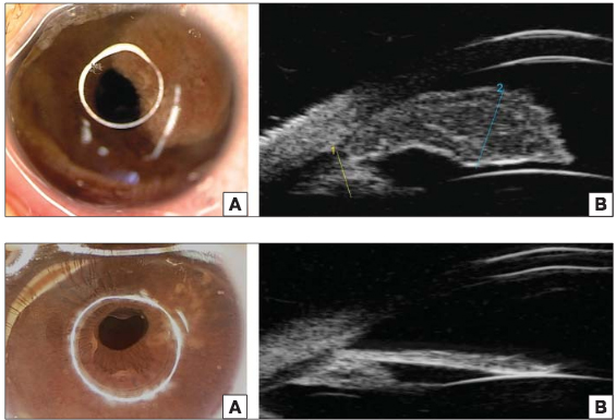
The infant was sent to Dr. Finger for evaluation of a spot on her iris. Slit-lamp examination revealed a left iris tumor that obscured the iris from pupillary margin to ciliary body, as you can see in the top two panels of the image below. Further examination via ultrasound biomicroscopy (UBM) led to a diagnosis of juvenile xanthogranuloma, which is a benign histiocytic skin disorder that primarily affects infants and children.

Dr. Finger treated the tumor with an injection of 20 mg of long acting sub Tenon’s corticosteroid followed by administration of topical steroid ointment four times per day. Within two weeks of the injection, the tumor had diminished to a flat whitish scar. Four and a half months after treatment began, there was no residual tumor visible, as shown in the bottom panels of this photo:
Upload: January 26, 2016

By publishing the results in the Russian Ophthalmological Journal, Dr. Finger’s successful treatment can be replicated to help patients across the Russophone world.

Few clinics engage in this level of international collaboration. Dr. Finger and his colleagues at NYECC are passionate about improving eye cancer diagnosis and treatment not just in North America, but around the world.


“Исследования Нью-Йорского Центра Онкологии Глаза Достигли Пациентов в России.

Доктор Фингер недавно опубликовал научную статью в Российском Офтальмологическом Журнале. Статья приводит описание клинического случая успешного диагноза и лечения 9-месячной девочки.

Этот клинический случай был уникален тем, что Доктор Фингер смог поставить диагноз и вылечить пациентку практически неинвазивным способом: без биопсии, и при применении кортикостероидов.

Новорожденная девочка была направлена к Доктору Фингер на осмотр пятна на радужной оболочке глаза. Осмотр выявил опухоль, распространившуюся от края зрачка до цилиарного тела, как видно на верхней половине снимка. Осмотр ультразвуковым биомикроскопом привел к диагнозу ювенильной ксантогранулемы. Это доброкачественная опухоль кожи, встречающаяся в основном у детей раннего возраста.

Доктор Фингер вылечил опухоль при помощи 20 миллиграмм локальной субэписклеральной инъекции кортикостероида и стероидной мази, применяемой четыре раза в день. В течении последующих двух недель, опухоль превратилась плоский белесый шрам. Четыре с половиной месяца после начала лечения опухоли не стало видно, как продемонстрировано на нижней половине снимка.

Публикация результатов этого успешного лечения, проведенного Доктором Фингер, в Российском Офтальмологическом Журнале позволяет так же успешно вылечить подобных пациентов в русско-язычных странах.

Немногие клиники в мире могут продемонстрировать такой уровень международного сотрудничества. Доктор Фингер и его коллеги в Нью-Йорском Центре Онкологии Глаза всегда стремятся улучшить диагностику и лечение опухолевых заболеваний глаза не только в Северной Америке, но и во всем мире.”

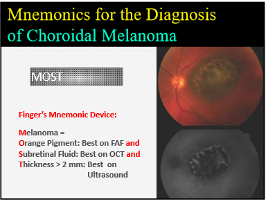
To Diagnose Choroidal Melanoma, Just Remember MOST

Dr. Paul Finger has developed a memory tool to help ophthalmologists identify and catch eye cancers. This simple but effective mnemonic serves as a guide to aide in knowing when to send a patient to an eye cancer specialist.

To effectively diagnose Choroidal Melanoma, ophthalmologists just need to remember “MOST.”

Upload: February 17, 2016

Dr. Tero Kivela presented Dr. Finger’s diagnosis methodology during a presentation at the 2016 World Ophthalmology Congress in Guadalajara, Mexico. Dr. Kivela serves as Director of the Ocular Oncology Service and Ophthalmic Pathology Laboratory at Helsinki University Central Hospital, and also teaches at the university. Dr. Finger was pleased to have Dr. Kivela present this work at the conference in his absence.

The presentation was designed to help participants recognize characteristics that differentiate benign from potentially malignant intraocular tumors of the uvea using up-to-date methodology. Dr. Finger’s MOST mnemonic makes up an important part of the methodology.

Small melanomas are difficult to diagnosis because little tumors have fewer easily identifiable characteristics. MOST provides an easy way for ophthalmologists to remember the key things to look for. It is generally accepted that the following three characteristics when seen together associated with a small pigmented choroidal tumor are diagnostic.

  1. Orange pigmentation on the tumor’s surface
  2. Leaking fluid around the tumor under the retina
  3. A thickness of 2 or more millimeters

So, physicians should remember that most tumors are caught with MOST:

Melanoma=

Orange pigment

Subretinal Fluid and

Thickness greater than 2 mm

Patients may also present with symptoms including: low visual acuity, shadow, metamorphopsia (distorted vision where straight lines in a grid appear wavy), photopsia (perceived flashes of light), and floaters. However, when they present with MOST, they are likely to have choroidal melanoma.

Following are the best ways to identify the three MOST characteristics.

O = A single wavelength of light is used to take a special intraocular photograph called fundus autofluorescence (FAF). This test offers the most effective means of identifying Orange pigmentation.

S = Optical Coherence Tomography (OCT) uses laser light to create a 3D image of the retina.  OCT is the best way to detect Subretinal fluid.

T = Ultrasound imaging is the “gold standard” for measuring intraocular tumor Thickness.

At the World Ophthalmology Congress, Dr. Kivela presented a study finding that 13 patients (8%) developed melanoma from a previously identified presumed nevus. Six of those patients had orange pigment, five had subretinal fluid, and three of the tumors were over 2mm in height.

Dr. Finger created MOST to help general ophthalmologists and retinal specialists in their decisions about who to refer for consultation.While MOST does not encompass all of the characteristics of choroidal melanoma, it is useful to distinguish most small melanomas.


Patient Stories

"Very well treated by Dr. Finger. He explained everything I needed to know about my issue with detail and attention, putting me at ease and giving me confidence to handle this problem for the rest of my life.”
N.N.

Read More

REQUEST AN APPOINTMENT

Go to Appointment Form

CONNECT AND SHARE