Second Eye Cancer Working Day Overview and Booklet

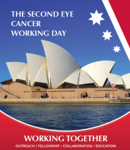
The Second Eye Cancer Working Day will take place Friday, March 24, at the International Convention Centre in Sydney, Australiaworking day book, at the Cookle Bay Room 1.

The Working Day provides an opportunity for eye cancer specialists from around the world to work together, face-to-face. The day will be divided into several sections, each dealing with a critical problem facing the specialty. Sections begin with an overview presentation from the moderators, followed by an interactive group session to brainstorm, complete work, and plan for the next meeting.

The 2017 Working Day will feature five separate committees focused on evidence-based medicine, educational programs, outreach to underserved areas, and multicenter quality assurance.

Click here for details about each of these committees and the physicians leading them.

If you can’t attend the working day, or if you are attending and want to get a preview of the day’s events, you can read the Second Working Day booklet here.

Click Here to download the booklet.


The Basics of Ophthalmic Ultrasonography (Video)

Ultrasound imaging is an effective method to visualize tumors on, within, and behind the eye. Ocular ultrasound machines generate sound waves that are reflected back by tissue in its path. The sound waves are then translated (by a computer) into an image.

Dr. Paul Finger has helped pioneer the use of ophthalmic ultrasound imaging in the diagnosis and treatment of eye cancers. In the following video, he presents the basic skills needed to perform ophthalmic ultrasound imaging. This includes the history of the evolution of these techniques as well as The New York Eye Cancer Center’s preferred practice patterns for evaluation of cancers of the eye and orbit.

Dr. Finger and his associates first described the use of 3D ultrasound for measurement of choroidal melanomas to document proper radioactive plaque placement and for extrascleral tumor extension. They also pioneered the use of computerized coronal C-scan ultrasound sections to measure the orbital portion of the optic nerve, to detect optic nerve sheath meningiomas and invasion of retinoblastoma into the optic nerve. Dr. Finger and his colleagues helped establish the use of high-frequency ultrasound (UBM) imaging for anterior segment tumors (melanocytoma, melanoma, cysts), and Dr. Finger has written on the use of high-frequency ultrasound for the diagnosis of conjunctival squamous carcinoma, periocular oncocytoma, Rosai Dorfman Disease, ciliary body adenocarcinoma and eyelid tumors.

This is important because high frequency ultrasound imaging is usually the best way to detect retinoblastoma hidden behind the iris, invasion of conjunctival tumors into the eye, and for measurement of iris and ciliary body tumors.

“High frequency ultrasound imaging has enabled eye cancer specialists to accurately measure anterior – iris and ciliary body – tumors, which in turn allows for eye and vision-sparing plaque radiation therapy,”Dr. Finger said.

This video not only provides a good overview of the techniques involved in ophthalmic ultrasonography, it will also point you to other resources where you can learn more.

“You have to know a little bit about ultrasound physics to be a great ophthalmic ultrasonographer.  You have to understand why you’re seeing what you’re seeing on the screen,” Dr. Finger said.

For more information contact us here.


2017 Eye Cancer Working Day Announcement and Schedule

International Society in Ocular Oncology and The Eye Cancer Foundation will sponsor the Second Eye Cancer Working Day on the first day of the ISOO meeting, Friday, March 24, at the International Convention Centre in Sydney, Australia, at the Cookle Bay Room 1.

The Working Day provides an opportunity for eye cancer specialists from around the world to work together, face-to-face. Our goal is to help the subspecialty working daymove forward into the mainstream of oncological care.  This will require the creation of evidence-based medicine, educational programs, outreach to underserved areas, and multicenter quality assurance.

The 2017 Working Day will feature five separate committees focused on these ongoing initiatives. These include the topics of international medical evidence, retinoblastoma fellowships, quality assurance, surgical standards, and consensus guidelines.

  • MIR: Multicenter International Registries create statistically significant evidence. These registries will improve patient care and help us defend our methods of diagnosis and treatment.
  • FOR- RB: Retinoblastoma fellowship initiative to address the worldwide RB mortality.
  • DRO: Quality assurance through Doctors Reporting Outcomes. Eye cancer specialists cannot know how to improve, unless they know the outcomes of their work.
  • COAST: A Comprehensive, open-access, consensus-based surgery text.
  • RASER: A prospective ophthalmic Radiation Side Effects Registry

The FIRST Working Day was held at The Curie Institute in Paris immediately prior to the ISOO 2015, and it was a big success.

We are excited to have the SECOND Working Day integrated with the biannual ISOO meeting. If you’re an eye cancer specialist attending the conference, be sure to mark your calendars and arrive by Thursday night!

Second Eye Cancer Working Day Schedule

Time: 8:00am – 5:00pm

Room: Cookle Bay Room 1, International Convention Centre

Convenors: Paul T Finger, Santosh G Honavar

Time Project
8:00am Registration and Coffee
8:30am – 9:00am


8:30am – 8:45am

8:45am – 9:00am

Introduction


Paul T Finger

Santosh G Honavar


9:00am – 10:00am


Comprehensive Open Access Surgical Textbook (COAST)

Convenor: Santosh G Honavar


Faculty: Fairooz P Manjandavida, Carol Shields, Zeynel Karcioglu, Mandeep Sagoo, Paul T Finger, Santosh G Honavar, Hardeep Mudhar, Sonal S Chaugule


10:00am – 11:00am


Radiation Side Effect Registry (RASER)

Convenor: Wolfgang Sauerwein


Faculty: Wolfgang Sauerwein, Paul T Finger, Brenda Gallie


11:00am – 11:30am


Morning Tea


11:30am – 12:30pm


Fellowship Outreach Retinoblastoma (FOR-RB)

Convenor: Ashwin Mallipatna


Faculty: Ashwin Mallipatna, Helen Dimaras, Brenda Gallie, Guillermo Chantada, James Muecke, Nathalie Cassoux, Santosh Honavar, John Zhao, Yacoub Yousef, Peter Gabel


12:30pm – 1:30pm Lunch
 

1:30pm – 2:30pm


Doctor Reported Outcomes (DRO)

Convenor: Tero Kivelä


Faculty: Tero Kivelä and faculty


2:30pm – 3:30pm


Multicenter International Registries (MIR)

Convenor: Bita Esmaeli


Faculty: Bita Esmaeli, Brenda Gallie, Martine Jager, Zeynel Karcioglu, Yulia Gavrylyuk, Paul T Finger


3:30pm- 4:00pm Afternoon Tea
4:00pm – 5:00pm Future Directions


Faculty: Santosh G Honavar, Martine Jager, Bita Esmaeli, Tero Kivelä, Ashwin Mallipatna, Wolfgang Sauerwein, Paul T Finger


**Please note that The ISOO Working Day workshop will be using live polling. Please ensure that you bring your mobile phone so that you can be an active part of the session.

Applying Chemotherapy Eye Drops Is Easier than It Sounds (Video)

When people hear the word chemotherapy, they generally think of intravenous medication injections to treat the whole body.  Of course, there are also chemotherapy drugs that can be administered orally in pill or liquid form. But there is another option available for treating eye cancer.

In 1993, Dr. Paul Finger and coworkers first reported on the use of Mitomycin chemotherapy eye drops to treat melanoma of the conjunctiva and cornea. Then in 2008, Dr. Finger and coworkers introduced interferon eye drops for extraocular melanoma. He realized that by placing chemotherapy agents directly onto the eye using drops, a high concentration of drug goes directly to the tumor. Topical treatment also limits the possibility of systemic (whole body) side effects.

Applying chemotherapy drops may sound complicated or intimidating, but it’s really not. In fact, most people put the drops in themselves.

This short video demonstrates the simple procedure.

As you watch the video, note the position of the patient. You also want to make sure excess medication does not get on your skin.

This video provides general information about applying chemotherapy eye drop, but as with any treatment, make sure you talk with your doctor about any questions. Not all chemotherapy eye drops are the same.

For more information contact us here.


What to Expect During Ophthalmic Plaque Radiation Therapy (Video)

Undergoing eye cancer treatment can be a scary prospect. At The New York Eye Cancer Center, we are committed to giving patients resources to make the experience as comfortable and stress-free as possible.

One way to do that is providing as much information as possible before treatment even begins. The following video shows one couple’s perspective on home-care during ophthalmic plaque radiation therapy.

EyePlaquePatientExperience from Paul T Finger on Vimeo.

Plaque radiation therapy is the most widely used treatment for choroidal melanoma and delivers a highly concentrated radiation dose to the tumor. This treatment results in relatively less radiation to surrounding healthy tissues.

Rice-sized radioactive seeds are attached within a gold or steel bowl called a plaque. This is surgically implanted to the wall of the eye, covering the base of the intraocular tumor. After surgery, the patient is typically sent home for five to seven days while the low-energy radiation sterilizes the tumor.

Dr. Finger performed plaque radiation treatment on Stanley Fishman.

“When I was first diagnosed diffuse iris melanoma, it was a rather frightening and daunting experience. Fortunately, Dr. Finger was able to guide me through the process very calmly and in a low-key fashion throughout all of the subsequent treatments and recovery.”

Now you can benefit from Stanley’s experience in this video featuring him and his wife. It provides some valuable insight on what to expect during the treatment period and also offers great tips on how to manage home care during this time.


Patient Stories

"Very well treated by Dr. Finger. He explained everything I needed to know about my issue with detail and attention, putting me at ease and giving me confidence to handle this problem for the rest of my life.”
N.N.

Read More

REQUEST AN APPOINTMENT

Go to Appointment Form

CONNECT AND SHARE