Eye Cancer Cluster in Raleigh, North Carolina

A recent article was just published in an online magazine called “Southerly” about a possible correlation between cancer incidence in Huntersville, North Carolina and industrial pollution. This relationship was scrutinized upon learning that several high school kids in a school by Lake Norman were diagnosed with ocular cancer. This prompted the residents to look for a correlation, and their current findings were found to be interesting. So far, there have been 20 cases of ocular melanoma in Huntersville within a population of 56 thousand people. Usually, ocular melanoma is found in 6 per million people. 

Soon, suspicions rose that the industrial pollution below the town could be related to energy development. However, finding a definitive cause for the incidence of cancer across many younger individuals proved more than difficult. Nevertheless, initiatives to fund research started outpouring from citizens. Doctors from Columbia University started to run tests on tumors of patients while the Huntersville Mayor has asked the town for help in funding more research. This research would show whether coal ash or heavy metals could be tied to these cancer clusters.          

In fact, there are many sites around North Carolina that are Superfund sites or industrial facilities, which are required by federal and state governments to test for pollutants. However, there are currently no standards for this testing when communities around the sites begin to develop schools, commercial buildings, and eventually neighborhoods. 

Nonetheless, it is clear that this town must discover answers to this heartbreaking incidence among high school children.


Melanoma 101: How Skin Cancer Can Impact the Eye

Right alongside cancers of the breast and lung, skin cancer exists as a well-known cancer afflicting U.S. Americans. The history of cancer study has lead to the identification of over 200 types of the disease, and skin cancer is the most commonly diagnosed of them, affecting more than 1 million Americans a year. Information on this type of cancer has been widely disseminated to the American public, from a fleet of dedicated websites, to news articles, and more, to the extent that most American adults realize that skin cancer can often arise from dangerous exposure to ultraviolet (UV) sunlight. But did you know that skin cancer and eye cancer, a lesser-known type of cancer, are closely linked? Indeed, skin cancer can negatively affect the eyes — take seventy-year-old John McPartland for example, who understood this well.

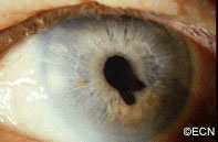
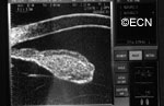
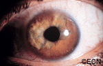
In 2001, McPartland, a lifetime lover of outdoor activity, noticed a freckle on his eyelid. Determined to find answers, he consulted with many doctors until finally meeting with Dr. Paul T. Finger, who diagnosed McPartland with conjunctival melanoma. A melanoma is a particular type of skin cancer; it affects nearly 70,000 people, and is found on the melanocyte cells of the skin. Melanocyte cells are responsible for the production of brown skin pigment — melanin, Because these pigment-producing cells are afflicted, melanomas commonly begin as pigmented, odd-looking freckles like McPartland’s. The conjunctiva is a delicate, clear membrane covering the inside of the eyelids and the white (sclera) of the eye. McPartland’s diagnosis was deadly.

“I just thought I should check it out and see if it is anything,” McPartland said, “and fortunately I did … As far as I’m concerned, [Dr. Finger] saved my life.”

About 2,400 patients are diagnosed with conjunctival melanoma every year, often in part due to the same UV light that causes skin damage. Those who work outdoors, play sports and/or frequent beaches are most vulnerable to eye cancer. Having light blue eyes and a fair complexion increases vulnerability, due to a lack of melanin (brown pigment) production that protects us from harmful sun exposure. Those who have a family or personal history of skin cancer are also vulnerable. For these people, Dr. Finger recommends that they “should have an eye exam, and then every six months thereafter.”

“Certain drugs also increase UV toxicity” Dr. Finger additionally cautions. “Patients who take chlorothiazides, sulfonamides, tetracycline, phenothiazins, psoralens, and allopurinol should be extra cautious about sunlight.”

A 2008 Fox News article highlights Dr. Finger’s experience with McPartland, along with his advice for optimizing eye health that he continues to recommend to his patients today. While the importance of wearing hats and using sunblock to protect the skin has long been stressed to the American public, Dr. Finger says people should approach this thinking to the eyes as well. The best way one can optimize eye health on their own? Using UV-blocking sunglasses!

“Think of sunglasses as sunblock for your eyes”, Dr. Finger says. He advises that sunglasses with 100% UV protection offer optimal prevention of sun damage to the eye.


Cancer is certainly a difficult reality to endure for many people, but there are ways one can help to protect themselves against the disease. By doing something as simple as wearing UV-blocking sunglass, you can take charge of your health today.

Stay tuned for more stories at eyecancer.com by keeping our website in your bookmarks!


Iris Melanoma

By Paul T. Finger, MD

Description

The iris is the colored part of the eye. It is made up of two layers. The outer “stroma” can be blue, hazel, green or brown. The back layer (the iris pigment epithelium) is always brown. Tumors can grow within, through and thus behind the iris.

Symptoms

This tapioca-colored iris tumor has pulled the iris pigment epithelium out onto the surface of the iris stroma.

Iris melanoma patients usually have no symptoms. The tumor might be noticed by the patient, their family, or by the eye care specialist (during a routine eye examination). Some people have lots of freckles on their irides. Some of these pigmented spots have thickness and are called Nevi.

If the patient notices that one of their nevi has changed, enlarged or is pulling (ovalization) on the pupil; they should see an eye care specialist for evaluation and referral to an eye cancer specialist.

Diagnosis

Photographs of the surface of the iris tumor should be obtained to establish a baseline for

Ultrasound shows diffuse iris thickening of the iris stroma with bowing and invasion of the underlying iris pigment epithelium.

future comparisons. High frequency ultrasound is used to examine and measure the iris tumor. Ultrasound can reveal if the tumor is cystic or solid, how it extends within the iris and ciliary body. Ophthalmic oncologists use high frequency ultrasound measurements to evaluate iris tumors for evidence of growth or regression after treatment.

Characteristics that suggest that an iris tumor is cancerous include seeing blood vessels within the tumor (intrinsic vascularity), secondary glaucoma, evidence that the pupil is deformed (ectropion uveae), and the development of a cataract beneath the tumor. Some eyes may have enlarged “sentinel”  blood vessels on the white of the eye (sclera) in the quadrant of the tumor.

The most important finding is documented growth. Since iris melanomas are commonly small, and less commonly (10-11%) spread to other parts of the body, these tumors are often watched for evidence of growth or change before biopsy or treatment is considered. A small amount of growth is not thought to significantly affect the rate of metastasis from a small iris melanoma. When necessary, eye cancer specialists can biopsy an iris tumor to help determine if the tumor is benign or malignant.

A diffuse iris melanoma causing severe glaucoma was too large to remove or irradiate and was treated by enucleation.
A diffuse iris melanoma causing severe glaucoma was treated by enucleation. At The New York Eye Cancer Center, we usually treat these cancers with eye and vision-sparing plaque radiation.

Treatments

Most pigmented iris tumors do not grow. They are photographed and monitored with periodic observation. When an iris melanoma is documented to grow, we know that it can damage the eye, cause secondary glaucoma and spread to other parts of the body. Then treatment risks become more acceptable to the patient. At The New York Eye Cancer Center, we typically use plaque radiation to destroy iris and iridociliary melanomas. This treatment allows for preservation of the iris, the pupil and does not cloud the cornea. We have found that the most common complication is cataract and permanent vision loss is rare.

Small Iris Melanomas:

Though most small iris melanomas can be surgically removed, however iridectomy may cause glare and astigmatism. The function of the iris and size of the pupil are better preserved if the tumor is destroyed with plaque radiation.

Medium-sized Iris Melanomas:

Though many of these tumors can be surgically removed, plaque or proton radiotherapy should  be considered as primary treatment for these tumors. Though a radiation cataract is likely to develop, due to the distance between the radiation and the macular retina, vision limiting radiation retinopathy is very unlikely.

Large-sized Iris Melanomas with Advanced Glaucoma:

These cases can often be difficult to treat with either surgical removal or eye-sparing radiation therapy. Cure for these tumors is likely to require removal of the eye.

Diffuse Iris Melanomas:

Sometimes the entire iris is filled with melanoma. In these cases, removal of the eye is a reasonable option. However, there has been a recent trend towards and our experience that eye and vision-sparing radiation of the entire front of the eye (anterior segment) can be used to control the tumor, spare vision and allow the patient to keep their eye.

Related links


Patient Stories

"Very well treated by Dr. Finger. He explained everything I needed to know about my issue with detail and attention, putting me at ease and giving me confidence to handle this problem for the rest of my life.”
N.N.

Read More

REQUEST AN APPOINTMENT

Go to Appointment Form

CONNECT AND SHARE