A New Support Group for Eye Cancer Patients is Available!

Stress and anxiety following treatment for choroidal melanoma have been well recognized among patients and studied among doctors. In fact, The NIH-funded Collaborative Ocular Melanoma Study reported on 209 patients with medium-sized melanoma treated with either brachytherapy or enucleation. In this sub-study, their goal was to compare the quality of life between treatment groups using questionnaires.

After questioning patients, researchers found that those undergoing radiation therapy had better quality of life outcomes related to their vision, such as driving, near activities, and binocular vision. After three to five years post-treatment, this benefit did decline, paralleling a decline in vision for the brachytherapy-treatment group (this, of course, predates the advent of vision-sparing anti-VEGF therapy).

However, in the scientific article published for the study, researchers state that “certain patients treated with brachytherapy, particularly those with pre-existing symptoms of anxiety, may suffer from increased risk of anxiety as compared with patients treated with enucleation during follow-up (Archives of Ophthalmology).”

At The New York Eye Cancer Center, we are currently participating in a study evaluating patient reported outcomes after plaque brachytherapy for choroidal melanoma. The more we understand a patient’s reception of plaque brachytherapy and the effect their treatment has had on their lives, the more we can specialize our care for each individual. We strive to offer compassion and understand what our patients are going through on a personal and psychological level. In an effort to help patients deal with their stress and anxiety, The New York Eye Cancer Center has begun to host a support group specifically for eye cancer patients and survivors. Please join Karen Campbell, a Licensed Clinical Social Worker (LCSW), who will facilitate this group. Sponsored by The Eye Cancer Foundation, this group therapy session will be held on Friday, October 13, 2017 at 1:30 pm at The New York Eye Cancer Center. Join us to have your voice heard among peers who understand what you are going through!

For your convenience, please consider downloading this flyer for the Support Group that contains all necessary information. We will host more sessions in the future, so in order to stay tuned for announcements on upcoming dates, please check back on eyecancer.com regularly!


New Study Highlights Promising Treatment for Patients with Iris Melanoma

A new study by Dr. Sonal Chaugule and Dr. Paul Finger has identified a promising treatment for patients diagnosed with iris melanoma. This is a first study describing regression characteristics in published literature.

Uveal melanoma is the most common primary intraocular malignancy in adults. Iris melanoma is the rarest cancer in this family, making up only 2% to 3% of cases. However, recent studies have found that biopsy-proven iris melanomas can spread outside the eye in up to 11% of cases. These findingsIris melanoma support the treatment of iris melanomas.

In the past, most patients with melanoma of the iris were treated by removal of the tumor along with the surrounding iris. Though the tumor is removed, the surgical procedure usually leaves a giant pupil with a non-functional iris sphincter and symptoms of glare.

“After part of the iris is removed, it is like having one pupil constantly dilated, even in the sun,” Dr. Finger said.

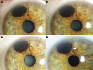
In their new study Chaugule and Dr. Finger describe patterns of tumor regression and side effects after iris-sparing treatment for iris melanoma using palladium-103 (103Pd) plaque brachytherapy. At the New York Eye Cancer Center, plaque brachytherapy has been found to be a conservative treatment modality with low local recurrence rate. Treatment with plaque radiation to sterilize the melanoma eliminates the need to open the eye, remove the tumor and make the pupil abnormally large.

Dr. Chaugule sought to examine and document the patterns of change after plaque radiation therapy for iris melanoma. The study included 50 patients with iris melanoma who underwent 103Pd plaque brachytherapy with at least 6 months of follow-up. Pre-treatment and post-treatment tumor morphology, gonioscopy and high frequency ultrasound imaging was studied and analysed.

In this study, palladium (103Pd) plaque brachytherapy of iris melanomas showed dissappearance of blood vessels within the tumor,, darkening of tumor surface, and decreased tumor thickness. With 100% local and systemic control at a mean duration of 5.2 years, the study shows this to be a safe and effective pupil sparing treatment.

You can click here to download and read the full study.


Iris Melanoma

By Paul T. Finger, MD

Description

The iris is the colored part of the eye. It is made up of two layers. The outer “stroma” can be blue, hazel, green or brown. The back layer (the iris pigment epithelium) is always brown. Tumors can grow within, through and thus behind the iris.

Symptoms

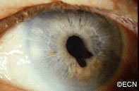
This tapioca-colored iris tumor has pulled the iris pigment epithelium out onto the surface of the iris stroma.

Iris melanoma patients usually have no symptoms. The tumor might be noticed by the patient, their family, or by the eye care specialist (during a routine eye examination). Some people have lots of freckles on their irides. Some of these pigmented spots have thickness and are called Nevi.

If the patient notices that one of their nevi has changed, enlarged or is pulling (ovalization) on the pupil; they should see an eye care specialist for evaluation and referral to an eye cancer specialist.

Diagnosis

Photographs of the surface of the iris tumor should be obtained to establish a baseline for

Ultrasound shows diffuse iris thickening of the iris stroma with bowing and invasion of the underlying iris pigment epithelium.

future comparisons. High frequency ultrasound is used to examine and measure the iris tumor. Ultrasound can reveal if the tumor is cystic or solid, how it extends within the iris and ciliary body. Ophthalmic oncologists use high frequency ultrasound measurements to evaluate iris tumors for evidence of growth or regression after treatment.

Characteristics that suggest that an iris tumor is cancerous include seeing blood vessels within the tumor (intrinsic vascularity), secondary glaucoma, evidence that the pupil is deformed (ectropion uveae), and the development of a cataract beneath the tumor. Some eyes may have enlarged “sentinel”  blood vessels on the white of the eye (sclera) in the quadrant of the tumor.

The most important finding is documented growth. Since iris melanomas are commonly small, and less commonly (10-11%) spread to other parts of the body, these tumors are often watched for evidence of growth or change before biopsy or treatment is considered. A small amount of growth is not thought to significantly affect the rate of metastasis from a small iris melanoma. When necessary, eye cancer specialists can biopsy an iris tumor to help determine if the tumor is benign or malignant.

A diffuse iris melanoma causing severe glaucoma was too large to remove or irradiate and was treated by enucleation.
A diffuse iris melanoma causing severe glaucoma was treated by enucleation. At The New York Eye Cancer Center, we usually treat these cancers with eye and vision-sparing plaque radiation.

Treatments

Most pigmented iris tumors do not grow. They are photographed and monitored with periodic observation. When an iris melanoma is documented to grow, we know that it can damage the eye, cause secondary glaucoma and spread to other parts of the body. Then treatment risks become more acceptable to the patient. At The New York Eye Cancer Center, we typically use plaque radiation to destroy iris and iridociliary melanomas. This treatment allows for preservation of the iris, the pupil and does not cloud the cornea. We have found that the most common complication is cataract and permanent vision loss is rare.

Small Iris Melanomas:

Though most small iris melanomas can be surgically removed, however iridectomy may cause glare and astigmatism. The function of the iris and size of the pupil are better preserved if the tumor is destroyed with plaque radiation.

Medium-sized Iris Melanomas:

Though many of these tumors can be surgically removed, plaque or proton radiotherapy should  be considered as primary treatment for these tumors. Though a radiation cataract is likely to develop, due to the distance between the radiation and the macular retina, vision limiting radiation retinopathy is very unlikely.

Large-sized Iris Melanomas with Advanced Glaucoma:

These cases can often be difficult to treat with either surgical removal or eye-sparing radiation therapy. Cure for these tumors is likely to require removal of the eye.

Diffuse Iris Melanomas:

Sometimes the entire iris is filled with melanoma. In these cases, removal of the eye is a reasonable option. However, there has been a recent trend towards and our experience that eye and vision-sparing radiation of the entire front of the eye (anterior segment) can be used to control the tumor, spare vision and allow the patient to keep their eye.

Related links


Patient Stories

"Very well treated by Dr. Finger. He explained everything I needed to know about my issue with detail and attention, putting me at ease and giving me confidence to handle this problem for the rest of my life.”
N.N.

Read More

REQUEST AN APPOINTMENT

Go to Appointment Form

CONNECT AND SHARE