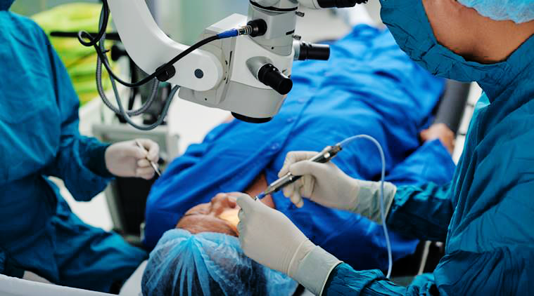
New Radiation Instructions for Eye Plaque Patients 2018
Based on published guidelines from United States Nuclear Regulatory Commission (NRC), The New York Eye Cancer Center and its affiliate New York Eye and Ear Infirmary of Mt. Sinai have agreed that patients undergoing low energy (iodine-125 or palladium-103) eye plaque radiation therapy for intraocular melanoma are allowed to proceed with their lives as usual.
Before this change, eye plaque patients were required to almost quarantine themselves. They had to remain at home, maintain a distance of 6-feet from others, and no pregnant women or children were allowed to visit.
With the new changes, patients can use public transportation. We ended most of the radiation exposure precautions and restrictions (i.e. you may go to the park, restaurants, grocery stores…etc.). However, it is recommended that patients stay at home as much as possible and that they do not engage in activities that could dislodge the implant and/or seeds. As before, the patient’s body fluids, clothing, and utensils ARE NOT radioactive and can be handled by others safely. We ask that the patient sleep alone and in a separate room away from anyone under the age of eighteen.
Dr. Finger says it’s about time: “With the radiation implants I use, eye cancer patients typically receive only a small fraction of the radiation given to patients undergoing implant radiation for other cancers, where patients are sent home on the day of implant.”
Though a few rules remain, they new radiation instructions are not nearly as strict. Free at last, patients can feel the sunlight, do their own shopping, and enjoy the company of their loved ones.
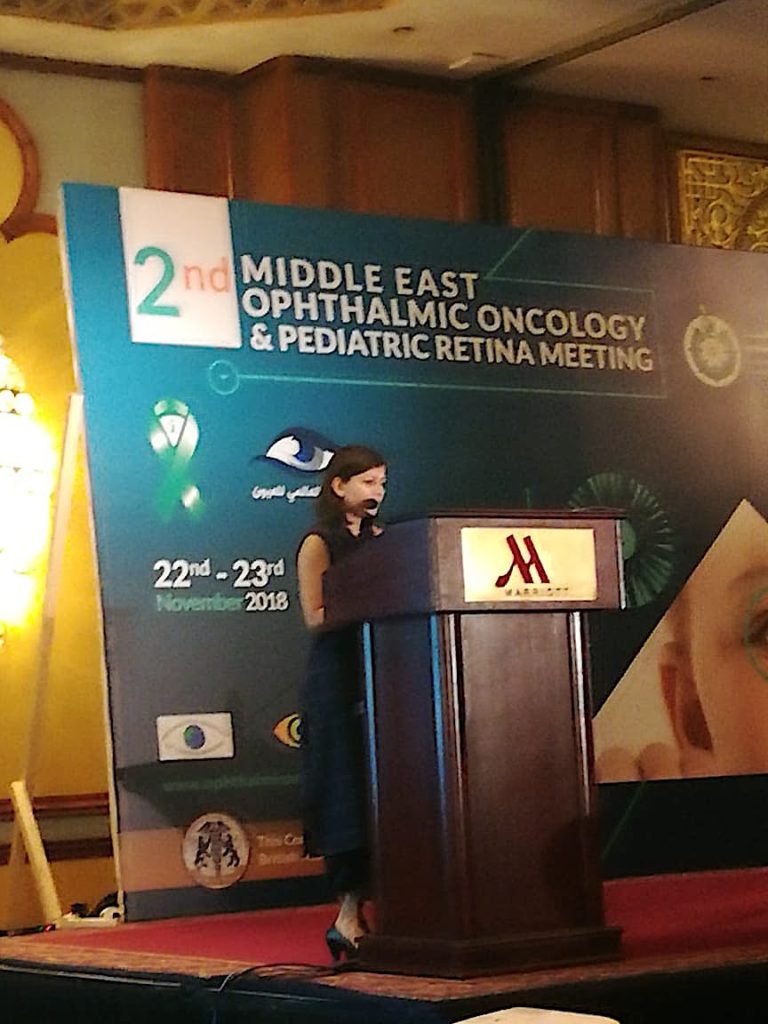
Dr. Abhilasha Maheshwari (above) presents ECF research at Ophthalmic Oncology Meet
November 22nd Cairo, Egypt:
November 22nd Cairo, Egypt: Physician scholars came together for The 2nd Middle East Ophthalmic Oncology & Pediatric Retina Meeting. Organized by Dr. Ihab Othman, the meeting fostered discussion of various topics in ocular oncology.
The Eye Cancer Foundation (ECF) fellow, Dr. Abhilasha Maheshwari, shared recent findings of ECF supported research. She first presented her research with her mentor and ECF-chair Dr. Paul T. Finger. The article, titled Regression Patterns of Choroidal Melanoma After Palladium-103 (103Pd) Plaque Brachytherapy, shows how choroidal melanomas regress after 103Pd plaque radiation therapy. Initially discovered as effective ocular cancer treatment by Dr. Finger in 1990, 103-Pd ophthalmic plaques have since been scientifically proven to be more gentle and effective than its precursor, 125-I. [See our results after 103Pd plaque therapy for choroidal melanomason our website!] Results showed the tumors became darker, decreased in thickness, and there was a reduction or complete elimination of the tumor vascularity.
Dr. Maheshwari continued her presentation on choroidal melanoma by discussing patients whose tumors were located very close to the optic nerve. This talk, “A 12-Year Study of Slotted Palladium-103 (103Pd) Radiation Therapy for Choroidal Melanoma: Near, Touching, or Surrounding the Optic Nerve, discusses how using specially designed plaques created by Dr. Finger, even melanomas that surround the optic nerve can be treated with eye and vision sparing radiation therapy (instead of removing the eye). Dr. Maheshwari’s results showed > 98% tumor control, while most had relative preservation their sight and eye.
The Eye Cancer Foundation takes great pride supporting research that improves patient care. Our fellows are given travel grants to spread the word around the world. As we spread knowledge of these unique findings and improved treatments, we also spread hope for those who would alternatively lose both sight and life.
To donate to the ECF and help sponsor fellowships, research, and cancer treatments, click here!
Byline: Published in The American Journal of Ophthalmology, 2019;198:45-53
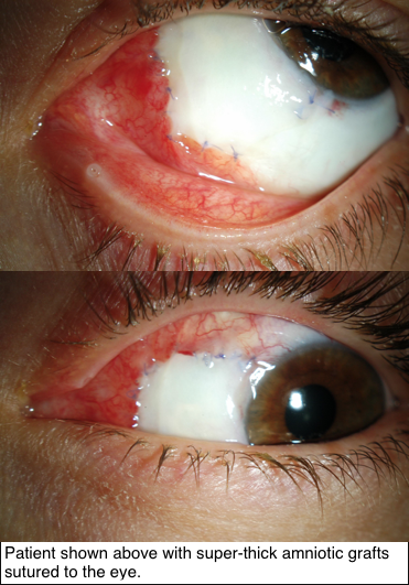
Since 1940, a single, thin layer of amniotic membrane graft (AMG) has often been used for repairing the cornea and conjunctiva. However, Dr. Finger says:
“our research shows that super-thick AMG (ST-AMG), up to ten times thicker than the prior AMG, is more effective for reconstruction of the eye’s surface.”
Research supported by The Eye Cancer Foundation has proved greater efficacy of this new technique in recreating the outer surface of the eye and inner surface of the eyelids. As published in the American Journal of Ophthalmology on November 2018, tumors of the conjunctiva and eyelids were surgically removed, then amniotic membranes from donor human placentas were sewn into the defects to recreate a normal ocular and inner eyelid surface.
Thus, amnion can provide a foundational platform for new cells to grow and flourish. In this case series, super thick amniotic membrane grafts (AMGs) were found to facilitate the healing of wounds.
How exactly does graft thickness affect the success of treatment? Well, the greater thickness means it is more easily sewn into the affected area, and
also helps the grafts to remain several weeks after placement. Thicker grafts are less likely to tear, rupture, or dissolve during the postoperative period. Most importantly, following treatment with ST-AMG, every single patient retained their sight and found their wounds successfully healed.
Super-thick amniotic membrane grafts have proven benefits to their thinner counterpart, and perhaps its versatility hints at potential for greater medical applications in the near future.
To keep up-to-date on the latest in Eye Cancer News, bookmark our website or follow us on facebook!
With a minuscule incidence of less than 0.005%, a myxoma is a staggeringly rare condition. Defined as noncancerous tumors of our connective tissue, myxomas present similarly to other, slightly more common conditions such as conjunctival lymphoma, lymphangioma, ocular surface squamous neoplasia (OSSN), or amelanotic melanoma. Consequently, they are usually misdiagnosed, or, at the very least, are difficult to diagnose.
Unique Ultrasound Findings!
Very limited literature exists describing cases and interventions of myxoma. In an effort to offset this shortage of research, The Eye Cancer Foundation sponsored a publication describing a myxoma case with unique ultrasonographic findings.
Other tumors of the conjunctiva often appear as a single, solid mass. Myxomas in particular present as smooth, yellow-pink masses on the eyeball, ranging in size from 4 mm to 20 mm. In this particular case under study, the patient’s myxoma showed scattered cells rather than a uniform image. So, while they are similar in many ways to other tumors, myxomas can have unique features that separate them from the others.
The Verdict…
Dr. Finger, Chairman of the ECF and Chief Researcher in this case study, concludes that:
“Though conjunctival myxomas can masquerade as various other conditions, high-frequency ultrasound proves myxomas have a distinct vascular pattern and no evidence of intraocular tumor invasion.”
October 2018–––In a recent publication in Cornea: The Journal of Cornea and External Disease, a noteworthy case report describes a patient with squamous cell carcinoma (SCC) in his only functioning eye who had his vision saved using a remarkable application of palladium-103 plaque radiation therapy.
Of all subtypes of conjunctival and corneal cancer, SCC is the most prevalent. The most common treatment for this type of superficial tumor is chemotherapy eye drops. However, in this particularly unique case, a functionally monocular patient presented with invasion of squamous cell cancer through a corneal wound into his eye. Maintenance of vision was essential for this patient, as he could only see using the affected eye.
Thus, a complex decision had to be made. Dr. Finger noted:
“Though we can treat even giant squamous carcinomas of the conjunctiva with topical chemotherapy eye drops, there is no evidence to support their use for intraocular tumor invasions. It is my opinion that it was unlikely that chemotherapy eye drops would penetrate deep enough into the eye to cure this patient.”
This was the potentially vision-saving observation. Since the SCC had spread internally, topical treatment was unlikely to reach the tumor. Had this treatment been chosen as a conservative intervention, the patient might have lost total vision and/or the entire eye.
In an effort to treat the tumor with minimum damage to the eye, palladium-103 plaque radiation therapy was chosen as an eye and vision-saving solution to the patient’s particular case. Such would simultaneously save the patient’s sight and their life.
According to the article, surgery was performed in June of 2014. Nearly 4 years later, the tumor has regressed without recurrence, and his vision is the same as before the radiation.
Thus, even in the face of the most unique cases, palladium-103 radiation therapy has proved remarkably effective.
For more information read the full case report here: [link]
The New York Eye and Ear Infirmary of Mt. Sinai Ocular Oncology Service [link]
The Fall 2018 issue of The Visionary is now available!
The Eye Cancer Foundation publishes The Visionary free-of-charge to keep you informed about the latest news, research, and global efforts focused on improving eye cancer treatment, diagnosis, and cure.
In this edition, you will find articles about:
Progress on the 20/20 Campaign, run by the ECF to provide worldwide retinoblastoma and eye cancer treatment.
A study providing evidence that even giant squamous melanomas can be cured using chemotherapy drops alone.
A mysterious cluster of eye cancer diagnoses within the graduates of the same university.
A personal insight from one of Dr. Finger’s long-standing patients.
Patients who have had treatment for a choroidal melanoma with radiation plaque brachytherapy will sometimes go on to develop retinal problems secondary to the radiation exposure which was needed to sterilize their tumor. This is called radiation retinopathy. These retinal problems can manifest as an increased thickness of the retina and/or intraretinal and subretinal fluid (fluid inside the retina and underneath the retina, respectively). If left untreated, this can lead to permanent loss of vision.
Fortunately, Dr. Finger had discovered that radiation retinopathy is treatable with periodic injections into the eye of anti-VEGF drugs, such as Avastin, Eylea, and Lucentis. These eye injections are already very commonly used to treat eye conditions, such as neovascular (“wet”) macular degeneration. In fact, the administration of these eye injections is now the most common surgical procedure in the United States.
There is now a new anti-VEGF drug called brolucizumab, also known as RTH258, which is currently in Phase III clinical trials. This novel drug is a tiny humanized single-chain antibody fragment. This type of fragment is thought to have improved tissue penetration, and therefore, improved drug delivery due to its small size. Trials thus far have shown brolucizumab to be superior in the treatment of wet macular degeneration, compared to Eylea. Patients presented with decreased retinal thickness, intraretinal fluid, and subretinal fluid — the same signs we treat in radiation retinopathy at The New York Eye Cancer Center! Interestingly, patients maintained these improved results even with a longer dosing interval of every 12 weeks. Most importantly, potential adverse reactions were comparable in both the brolucizumab and Eylea groups. Pending the results of more trials and FDA approval, this drug may be available for use in 2019.
To stay updated on all the latest eye cancer research, please keep our website, eyecancer.com in your bookmarks!
Retinoblastoma (RB) is the most common eye cancer in children, affecting approximately 8,000 of them each year. In developed countries like the United States, the survival rate reaches beyond an astounding 96%, with early diagnosis and treatment being key to saving a patient’s life and sight. However, this incidence rate is higher in developing countries, where most of the children succumb to metastatic retinoblastoma. In areas where children and families have no means of travel to treatment centers far away from them, these afflicted children often endure their disease untreated until there is very little hope for them left. Because no child or family should have to suffer these losses, especially due to the simple inability to reach proper care, The Eye Cancer Foundation has launched the 2020 Campaign, a campaign dedicated to training ophthalmic oncologists to serve in underprivileged countries.
One such underprivileged area is India, a country populated by over 1.32 billion people and counting, where 1,500 of the global 8,000 retinoblastoma cases are diagnosed every year. However, the reality persists that many cases of retinoblastoma go undetected or unreported in India, and awareness for the disease is abysmally low in rural areas. Motivated by India’s need to increase awareness and treatment for this disease, The Eye Cancer Foundation has sponsored fellowships for three doctors from India to train with Dr. Paul T. Finger at The New York Eye Cancer Center over the last year alone — Dr. Sonal S. Chaugule, Dr. Abhilasha Maheshwari, and Dr. Puneet Jain.
After the successful completion of her NYECC-ECF fellowship in Summer 2017, Dr. Chaugule returned to her native Maharashtra, India. She currently employs her expertise in retinoblastoma care by consulting at HV Desai Eye Hospital, a critical center for eye cancer patients in Pune, India. Her continued efforts to raise awareness in this vastly unrecognized disease have led to her medical advice being featured across Indian news media. According to Dr. Chaugule in The Indian Express, “Awareness about retinoblastoma is low and early detection is crucial to give the best chance of saving the child’s life, eye, and vision. Early detection and proper treatment will ensure 95% of the children diagnosed with RB are saved from death, 90% have their eye intact and 85% have their vision protected.
Unfortunately, in India, a child is taken to an eye specialist only when there is any notable problem, which makes treatment of RB at a later stage much harder,” she said.
Dr. Chaugule suggests that systemized screening of the eye for any abnormality in infants and toddlers should be made mandatory. Additionally, it is crucial that all doctors and healthcare professionals, whether they be eye cancer specialists or not, ought to be deeply sensitized to this disease’s magnitude.
In response to India’s growing need for retinoblastoma care, The Iksha Foundation, a non-governmental organization based in Benglauru, has accelerated their programs to raise awareness for the disease so that children may be diagnosed early enough to save their livees. Founder and trustee at the Iksha Foundation, Thanmaya Bekkalale, says, “We only know the reported cases of retinoblastoma — there are numerous cases that go unreported. The need of the hour is to spread individual and societal awareness about retinoblastoma and promote early detection as it is documented that every day, four children are born with eye cancer in India, and one of them is facing death as a result of diagnosis at an advanced stage, or not diagnosed at all.”
To raise awareness, May 13th through 19th were observed as World Retinoblastoma Awareness Week. The Iksha Foundation will hold awareness programs, ensuring that their various stakeholders will understand that early diagnosis is crucial to saving the lives of children throughout India.
Read the article published in The Indian Express by Dr. Chaugule and her colleagues at HV Desai Eye Hospital here.
To stay up-to-date on the latest news in eye cancer, please keep our website, eyecancer.com, in your bookmarks.
In our previous blog, we unveiled Dr. Finger’s Results page, the first public database of its kind to report a doctor’s treatment outcomes. With the power of the world-wide-web at our fingertips, it is now easier than ever to browse for healthcare options. Search engines, with the simple press of a button, are able to provide patients with a virtually infinite list of specialists available to them locally, regionally, even internationally.
So, shouldn’t it be just as easy to know how successful these specialists are? How can patients choose the best doctor without knowing their past performance? These questions motivated the creation of Dr. Finger’s Results page, a launch that was met with glowing approval from across both patient and scientific communities. And now, this page is more comprehensive than ever!
Understanding the Report
Choroidal melanoma, iris-ciliary body melanoma, and squamous conjunctival malignancy are three of the most common conditions treated at The New York Eye Cancer Center. Once treated for these select diseases (whether through plaque radiation, chemotherapy, cryotherapy, and so on), patients are routinely seen at NYECC in follow-up visits, where they are monitored for any changes in tumor activity and quality of life.
Starting from December 1, 2017, each NYECC patient seen in these follow-up visits is anonymously entered in our Result’s page database — information that becomes immediately accessible on our website. These results are updated weekly, and with Dr. Finger’s practice spanning over 30 years, the database will continue to grow moving forward.
For each disease, we report on:
– Patients Entered: The number of patients included in these results, which grows with every week once patients are seen by Dr. Finger in follow-up.
– Visual Acuity: The average and median (most common) visual acuity, or eye chart test score, after finishing treatment.
– Local Tumor Destruction: The percentage of patients whose tumors are successfully eliminated through treatment.
– Initial Eye Removal: The percentage of patients who have undergone enucleation (eye removal) surgery prior to being treated by Dr. Finger at NYECC.
– Metastases: The percentage of patients whose tumors have spread to other organs after treatment.
– Average Follow Up: Number of years after treatment before additional treatments are required.
The data, located on our Results page and observable through an interactive table, reports on patients treated only by Dr. Finger. Patient data is strictly confidential, HIPPA-compliant and, once again, anonymous.
What’s New
Our Results page has a new look! Rather than having to observe all reports at once, we have implemented a ‘choose your results’ feature. This cancer directory allows you to choose which of these three diseases you would like to observe. Choroidal Melanoma, Iris Ciliary Body Melanoma, Squamous Conjunctival Malignancy — each of these reports now has its own page and table. These pages will be a source of information specialized to each disease; the result is a streamlined, organized process for eye cancer patients across the world. Here is a shortcut to the directory you can find on our page:
The launch of our results page is the first step, and we encourage other centers to join us in this effort. The Eye Cancer Foundation will offer assistance to any center or solo practitioner in setting up a page akin to the new NYECC Results page.
Let’s hold ourselves accountable to our outcomes and empower patients to make their life-changing choice of eye cancer specialist based on visible results.
With 87,110 diagnoses estimated to be made in 2018 for U.S. Americans, skin cancer, particularly a melanoma, is the most frequently diagnosed cancer in the United States. Ocular melanomas, however, remain uncommonly diagnosed, affecting just six in every one million people a year. Given the extreme rarity of ocular melanomas, doctors and researchers were shocked to find this disease found in highly concentrated numbers in two states. A total of 36 people — all graduates from Auburn University, Alabama — were diagnosed with ocular melanoma. From Huntersville, North Carolina, 18 patients were also found out to have the same disease.
Left to right: Ashley McCrary, Allison Allred, Juleigh Green and Lori Lee
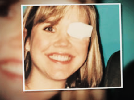
Juleigh Green, Allison Allred, and Ashley McCrary had spent their years at Auburn University as close friends. And later on in their lives, each woman discovered they had the same rare disease. Juleigh Green was the first among her friends to be diagnosed. Just 27 at the time, Green was experiencing strange flashes of light obstructing her vision and consulted her ophthalmologist immediately. In an interview with CBS, Green explains her shock upon what they found:
“[My doctor] said, ‘There’s a mass there, there’s something there, I don’t know what it is, but it looks like it could be, you know, a tumor,’” Green said. “It’s like you had the breath knocked out of you, you know?”
Allison Allred, who also was experiencing flashes of light for 7 to 10 days, was the second of their friend circle to be diagnosed in 2001, at the age of 31. Her doctor had first believed that the flashes were due to a retinal detachment. According to Allred, her doctor had said: “Well, [the retina] is detached because there’s a 10 millitmeter melanoma sitting on it.”
Both Green and Allred opted for enucleation and have had their afflicted eye removed. However, Allred’s extremely aggressive and stubborn cancer has since recurred nine times in six separate places in her body. “Two days ago I found out that it’s come back to my brain,” Allred told CBS, “So, I’m actually gonna have radiation on my brain tomorrow.”
Ashley McCracy was the third friend, with her own diagnosis coming from observing unusual black spots along her iris. In an interview with CBS News correspondent Anna Werner, McCrary said:
“What’s crazy is literally standing there, I was like, ‘Well, I know two people who’ve had this cancer.”
“And did you understand then how strange that was?” asked Werner.
“No. No, I didn’t.”
All three women were treated at Sidney Kimmel Cancer Center at Thomas Jefferson University in Philadelphia, Pennsylvania. McCrary mentioned Green and Allred’s similar diagnosis to her oncologist at Kimmel Center, Dr. Marlana Orloff.
Orloff was baffled.
“Most people don’t know anyone with this disease,” Orloff said. “We said, ‘OK, these girls were in this location, they were all definitively diagnosed with this very rare cancer — what’s going on?”
A fourth Auburn alumna, Lori Lee, is also being treated at Kimmel Center. “This is a rare cancer, so it’s not like you can just go anywhere and have anybody know anything really about it,” Lee said. “Until we get more research into this, then we’re not gonna get anywhere. We’ve got to have it so that we can start linking all of them together to try and find a cause, and then one day, hopefully, a cure.”
Orloff and her fellow researchers and oncologists at Kimmel Center immediately began to investigate this bizarre case. Thus far, the Alabama Department of Health states that “it would be premature to determine that a cancer cluster exists in the area”. Officials at Auburn University hope that research will help illuminate the cause of this rare cancer appearing at such high concentrations in Alabama and North Carolina.
Each patient’s emotional response to this mystery cannot be understated. “That was very hard for me,” McCrary told CBS. “Growing up, the one thing that I liked about myself was my eyes.”
Juleigh Green, pictured above, was the first of her two friends from Auburn University to be diagnosed with ocular melanoma.
McCrary’s personal journey in dealing with her cancer diagnosis lead to the creation of the Auburn University Ocular Melanoma Page on Facebook, which has astoundingly discovered 36 more graduates afflicted with ocular melanoma. The Facebook Page offers itself as an effective support network for these graduates.
Kimmel Center researchers continue to look for answers to explain these phenomena both in Alabama and North Carolina.
Stay tuned for the latest updates on this case and others by keeping eyecancer.com in your bookmarks.
"Very well treated by Dr. Finger. He explained everything I needed to know about my issue with detail and attention, putting me at ease and giving me confidence to handle this problem for the rest of my life.”
– N.N.