Every two years, eye cancer specialists worldwide convene at the International Society of Ocular Oncology Conference in order to review latest research and encourage international collaboration within the field of ophthalmic oncology.
For details on the ISOO Biennial Conference, read more here.
How Can You Tell a Choroidal Melanoma is Regressing?
Dr. Abhilasha Maheshwari, ECF fellow
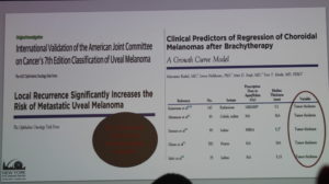
Dr. Abhilasha Maheshwari was the first Eye Cancer Foundation fellow to lecture today with her presentation on the paper Regression Patterns of Choroidal Melanoma after Plaque Brachytherapy, published alongside Dr. Paul T. Finger. In the two-minute rapid-fire session, Dr. Maheshwari filled us in on her data points from the study, concluding that there are various markers that show us when a choroidal melanoma is controlled after treatment with plaque brachytherapy. She provided us various markers, such as:
Dr. Finger and Dr. Maheshwari
Increased tumor pigmentation.
Ultrasonography results:
maintenance of dome/mushroom shape
decreased tumor thickness
increased internal reflectivity
Fundus autofluorescence imaging showed a pattern of increased followed by decreased organge pigment lipofuscin
OCT showed decreased exudative subretinal fluid, and resolved drusenoid retinal pigment epithelial detachments (DRPED)
Flurocein angiography showed resolution of intrinsic tumor vascularity
Each of these characteristics serve as indicators of tumor regression. Dr. Maheshwari gave us one final take-away message: better initial local control means extremely less systemic spread, which overall means better patient outcomes.
Dr. Finger and Dr. Maheshwari in the “hot seats” up on stage – Every new session rotates a panel of doctors who have given lectures in order to ask and answer questions and foster discussion.
Interesting New Finding: One talk updated us on recent clinical trials for a drug that showed promise in laboratory studies for reversal of the effect of BAP1-related tumor progression. Perhaps we will hear more on this at the next ISOO!
Unique Case Report: Two, Simultaneous Primary Melanomas
Dr. Maheshwari returned for her second talk of the day, presenting results from Bilateral Simultaneous Primary Choroidal Melanomas: Treated with Palladium-103 Plaque Radiation, published along with Dr. Paul Finger. She elaborated on an interesting case who presented with separate, unrelated choroidal melanomas, one in each eye. After sequential treatment to maximize outcomes, the patient remains at 20/25 vision and shows great outcomes since their latest follow-up.
Unique Case Report: 7 Years After Treatment of a Primary Melanoma, A Second Primary is Found
Dr. Finger gave his first talk of the event to present a unique case of a patient who had been successfully treated for a stage T1 ocular melanoma. Seven years after treatment, however, she developed choroidal metastasis (this one, T3). Following intensive immunotherapy, the patient showed tumor controlled and marked decrease in liver metastasis. Upon follow-up, the patient still sees 20/25 and is maintained with anti-VEGF therapy.
With this talk, Dr. Finger wants to emphasize “the importance of consistent, long-term, post-treatment periodic ophthalmic and systemic surveillance for patients with choroidal melanoma.”
Dr. Finger Presents a Unique Case!
Identifying Genes Associated with Cancer Predisposition
Dr. Colleen Cebulla presented interesting findings in her presentation, Whole exome sequencing to identify candidate genes associated with hereditary predisposition to uveal melanoma. She covered various, possibly cancer-related genes other than the well-known, BAP1 mutation. At the beginning stages of research, the project has already analyzed 29 cases and identified a significantly large incidence of the genes SMARCE1,, PALB2, and MLH1. Particularly of interest are PALB2, which has already been linked to breast, ovarian, and pancreatic cancer risk, and MLH1, which has been linked to Lynch syndrome and has been deemed responsible for multitudes of different tumors.
Dr. Cebulla is happy to welcome additions to the study and to be contacted if any centers are willing!
“SORTT” for Short: Study of Ophthalmic Radiation Therapy Toxicity
Unfortunately unable to attend, Dr. Wolfgang Sauerwein requested Dr. Finger to present on Study of Ophthalmic Radiation Therapy Toxicity (SORTT): a prospective international survey. A major talk of the day, SORTT proposes “a prospective, multicenter, international data registry that collects structured information on toxicities after radiotherapy for eye cancer. This will offer the medical evidence needed for ophthalmic radiation side effect staging and modality selection.” This data may also help to create an AI to help physicians select the optimal radiation modality for each individual case.
Another jam-packed session of presentations, lectures, research findings, and plans for greater multicenter cooperation. As Day 2 comes to a close, Day 3 promises more on melanoma, other intraocular cancers, and the beginning of retinoblastoma discussions.
In our previous blog, we unveiled Dr. Finger’s Results page, the first public database of its kind to report a doctor’s treatment outcomes. With the power of the world-wide-web at our fingertips, it is now easier than ever to browse for healthcare options. Search engines, with the simple press of a button, are able to provide patients with a virtually infinite list of specialists available to them locally, regionally, even internationally.
So, shouldn’t it be just as easy to know how successful these specialists are? How can patients choose the best doctor without knowing their past performance? These questions motivated the creation of Dr. Finger’s Results page, a launch that was met with glowing approval from across both patient and scientific communities. And now, this page is more comprehensive than ever!
Understanding the Report
Choroidal melanoma, iris-ciliary body melanoma, and squamous conjunctival malignancy are three of the most common conditions treated at The New York Eye Cancer Center. Once treated for these select diseases (whether through plaque radiation, chemotherapy, cryotherapy, and so on), patients are routinely seen at NYECC in follow-up visits, where they are monitored for any changes in tumor activity and quality of life.
Starting from December 1, 2017, each NYECC patient seen in these follow-up visits is anonymously entered in our Result’s page database — information that becomes immediately accessible on our website. These results are updated weekly, and with Dr. Finger’s practice spanning over 30 years, the database will continue to grow moving forward.
For each disease, we report on:
– Patients Entered: The number of patients included in these results, which grows with every week once patients are seen by Dr. Finger in follow-up.
– Visual Acuity: The average and median (most common) visual acuity, or eye chart test score, after finishing treatment.
– Local Tumor Destruction: The percentage of patients whose tumors are successfully eliminated through treatment.
– Initial Eye Removal: The percentage of patients who have undergone enucleation (eye removal) surgery prior to being treated by Dr. Finger at NYECC.
– Metastases: The percentage of patients whose tumors have spread to other organs after treatment.
– Average Follow Up: Number of years after treatment before additional treatments are required.
The data, located on our Results page and observable through an interactive table, reports on patients treated only by Dr. Finger. Patient data is strictly confidential, HIPPA-compliant and, once again, anonymous.
What’s New
Our Results page has a new look! Rather than having to observe all reports at once, we have implemented a ‘choose your results’ feature. This cancer directory allows you to choose which of these three diseases you would like to observe. Choroidal Melanoma, Iris Ciliary Body Melanoma, Squamous Conjunctival Malignancy — each of these reports now has its own page and table. These pages will be a source of information specialized to each disease; the result is a streamlined, organized process for eye cancer patients across the world. Here is a shortcut to the directory you can find on our page:
The launch of our results page is the first step, and we encourage other centers to join us in this effort. The Eye Cancer Foundation will offer assistance to any center or solo practitioner in setting up a page akin to the new NYECC Results page.
Let’s hold ourselves accountable to our outcomes and empower patients to make their life-changing choice of eye cancer specialist based on visible results.
The wall of the eye has 3 main layers. From outside to inside there is: the white sclera, a blood vessel layer called the uvea (choroid, ciliary body and iris) and an inner retinal layer. Further, the pigment producing cells, “melanocytes” are primarily found in the vascular uveal layer. It is those melanocytes that can turn into malignant melanoma. Therefore, when melanoma happens in the choroid, they are called “choroidal melanoma,” the most common primary intraocular malignancy in adults. That said, choroidal melanomas are rare with 5 to10 out of each million people diagnosed with a choroidal melanoma each year. Choroidal melanomas can spread to other parts of the body.
Eye cancer specialists can determine if you have a choroidal melanoma by performing a complete eye examination with testing. This includes asking questions about your medical history, examining both of your eyes, looking into the eye through a dilated pupil, performing an ultrasound examination, and specialized photography (to examine the circulation within the choroidal melanoma).
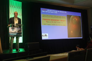
MOST – Fingers’ Melanoma Mnemonic
Dr. Finger has developed the mnemonic device “MOST” to help eye care specialists to determine if the intraocular tumor is a melanoma.
“M,” Melanoma:
“O,” Orange Pigment Lipofuscin (OPL) a metabolic side product of cell death. This finding tells us that that either the underlying tumor is destroying the overlying tissue or itself is degenerating. Lipofuscin is best seen with a photographic test called Fundus Auto Fluorescence imaging, or FAF.
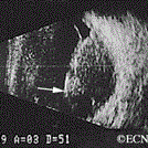
“S,” Subretinal fluid (SRF) is created by poorly formed or new, “neovascular” blood vessels within the tumor. Cancers need new vessels in order to grow. Large amounts of SRF can be seen by ophthalmoscopy (looking into the eye) and ultrasound imaging. However, small amounts of SRF are best seen on 3D optical coherence tomographic imaging (3D-OCT).
“T,” Thickness of the tumor has been associated with malignancy. Simply, the thicker it is the more likely a pigmented intraocular tumor is malignant. It is widely accepted that tumors greater than 2.0 mm are more likely to be malignant. Ultrasound imaging is currently the best method to measure tumor thickness.
Your specialist will also request that you have a complete general medical check up and specific tests depending upon what they see inside your eye. In the Collaborative Ocular Melanoma Study (COMS), participating eye cancer specialists correctly diagnosed select choroidal melanoma in over 99.6% of cases (without a biopsy). That said, patients with unusual appearing “atypical” tumors were not entered into the study.
Classic Indications for Biopsy
Atypical tumor, metastatic tumor with no observable primary cancer and when the patient requests a pathology diagnosis. More recently, primarily due to genetic testing services, more and more centers are routinely performing choroidal tumor biopsy primarily for genetic tumor analysis. Genetics offers information about the tumor, but does not allow doctors to avoid treatment or follow up systemic testing for metastasis.
Choroidal biopsy has been associated with a risk of hemorrhage, infection, retinal detachment and a poorly quantified risk of tumor seeding (outside the eye). Risks related to tumor seeding are thought to be small, but clearly they have not been evaluated by any large prospective or retrospective study. Each eye cancer specialist should discuss the relative risks (known and unknown) of biopsy prior to surgery.
Questions About Intraocular Biopsy:
Remove the need for surgical tumor treatment?
Reduce the number of radiologic examination or years needed for systemic surveillance?
What are the risks of biopsy (hemorrhage (e.g. vitreous, subretinal, subfoveal), seeding, damage to the lens, optic nerve, retinal detachment, cataract, epiretinal membrane, loss of vision, loss of eye and/or reaction to anesthesia).
Symptoms
Most choroidal melanoma patients have no symptoms. The melanoma is found on routine eye examination. If patients have choroidal melanoma symptoms, they are usually seeing “flashes of light,” noticing “distortion” or loss of vision, and floating objects (floaters) in their vision.
If the choroidal melanoma is in the front of the eye (near the natural lens), it can push or tilt the natural lens causing an irregular astigmatism (blurring of vision).
Choroidal melanoma can leak fluid beneath the retina, making the retina detach and cause symptoms of flashing lights and floating specks “floaters”.
If the choroidal melanoma is in the macula (center of vision), it can grow beneath the fovea making the patient far-sighted. The choroidal melanoma can also grow into and destroy the fovea causing distortion, loss of vision or changes in color perception.
It is important to note that most patients with choroidal melanoma have no symptoms at all. Their tumors are found when they visit their eye doctor for a “routine” eye examination. So everyone should have periodic eye examinations (including dilated ophthalmoscopy). In general, the earlier and smaller the choroidal melanoma, the better the prognosis for both life and sight.
Other, more unusual presentations of anterior choroidal and iridociliary melanoma are discoloration of the iris, a brown spot on the outside of the eye, an irregularly shaped pupil and glaucoma.
Diagnosis
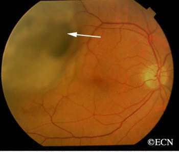
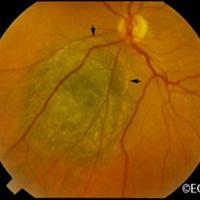
Choroidal melanoma is usually seen by ophthalmoscopy (when your eye doctor looks through a lens into your dilated pupil). Choroidal melanoma has typical “diagnostic” characteristics that include but are not limited to: pigmentation, low to moderate internal ultrasound reflectivity, clumps of orange pigment lipofuscin on its surface, leakage of subretinal fluid, or retinal detachment (on or around the choroidal melanoma) and thickness.
Pigmentation is due to naturally occurring melanin that comes from melanocyte cells in the choroidal layer of the eye. Choroidal melanomas are most commonly pigmented, but can be variably pigmented and even non-pigmented (amelanotic). Non-pigmented choroidal melanoma is due to a proliferation of melanocytes that have lost their ability to make the melanin pigment.
Orange pigment is made up of a chemical called lipofuscin and appears on the surface of choroidal melanomas. Lipofuscin is a product of cell death which indicate that cells are dying on the tumor’s surface. This is also sign of metabolic activity. Melanomas are more metabolically active than choroidal nevi.
Ultrasound is typically used to measure the choroidal melanoma size, evaluate internal tumor reflectivity, and look for melanoma extension behind the eye into the orbit called extrascleral extension. Ultrasound imaging has demonstrated that most choroidal melanomas are shaped like a dome and less commonly like a mushroom. Ultrasound can also evaluate and detect choroidal melanoma associated retinal detachment. However, optical coherence tomography (OCT) is a more sensitive way to detect subretinal fluid – retinal detachment.
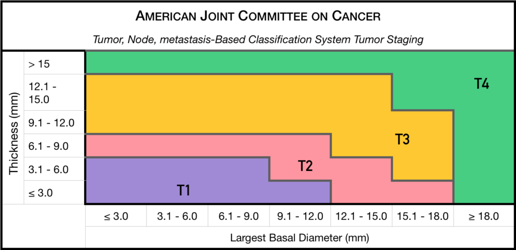
Staging
Chaired by Dr. Paul T. Finger, a committee of top ophthalmic specialists assembled to form the AJCC-UICC Ophthalmic Oncology Task Force. To ensure a broad range of specialists, Dr. Finger “internationalized” the committee, including over 58 members both from the USA and around the world.
This committee had one driving goal: to design a clinically useful Tumor-Node-Metastasis (TNM) based classification “language” for ocular tumors. This first-of-its-kind classification system has become a universal language for all who diagnose and treat ocular tumors.
Not only does a universal classification system offer cancer staging for the patient, it also allows physicians to directly compare data. In the long run, a common “eye tumor” language helps usdetermine and differentiate treatment types as well as coordinate the efforts of researchers working for a cure.
Dr. Finger has since translated this staging system for the worlds’ Union International for Cancer Control (UICC), and offered it to the world. In order to get everyone to employ this new language, he has recruited all the major medical journals to require eye cancer researchers to use AJCC-UICC staging.
Treatments
Small Choroidal Melanoma (AJCC T1 and T2): Patients with a small choroidal melanoma can be treated after their first visit, but since growth helps to prove that the tumor is a cancer, your doctor may suggest “observation” or watching for a small amount of choroidal melanoma growth prior to treatment. Your eye cancer specialist should discuss the relative risks and potential benefits of “observation for growth” as compared to “immediate treatment” for choroidal melanoma. If growth is documented (typically within 6 months of observation), eye cancer specialists will typically recommend definitive treatment.
Medium-sized Choroidal Melanoma (AJCC T3 and T4): Most patients with large-sized choroidal melanoma can be also be treated with eye-sparing low energy radiation therapy (e.g. palladium-103). However, larger tumors require more radiation and larger irradiated intraocular volumes resulting in greater risk of radiation side-effects and poor vision. Rarely such eyes have to be secondarily removed. Eye cancer specialists try to preserve eyes, even if the eye had reduced vision.
Large-sized Choroidal Melanoma: Very large choroidal melanomas (greater than 22 mm width) may be treated by initial removal of the eye (enucleation). This is because the amount of radiation required to destroy a choroidal melanoma that fills most of the eye will likely be too much for the eye to tolerate.
However, most patients, even with very large-sized choroidal melanoma can be treated with eye-sparing radiation therapy. However, after eye sparing radiation for very large choroidal melanomas, eyes are at greater risk to have poor vision, secondary inflammation and may require secondary removal at a later date.
Additional Info
Patients often ask why they have a choroidal melanoma. While there is no one cause, choroidal melanoma is more common among patients with blue vs. brown eyes, those with outdoor occupations, and in Australia where there is a hole in the ozone layer. Therefore, though this hypothesis has yet to be proven, it seems reasonable to assume that choroidal melanoma is related to sunlight (ultraviolet exposure).
In that sunlight exposure has been linked to several eye cancers and diseases of the eye, Dr. Finger suggests that you “Think of Sunglasses as Sun Block for your Eyes” ™ and start wearing your UV blocking sunglasses. They make great gifts too!
Dr. Finger also often gets questions related to stage and spread. These two things are closely linked––choroidal melanoma size is most closely related to its risk for spread to other parts of the body (metastasis). In three separate studies, cumulatively involving almost 20,000 patients, the average rate of metastasis has been 50%. However, patients with smaller tumors have much lower rates compared to larger tumors. Therefore, patients should ask their eye cancer specialist about their tumors AJCC-UICC tumor size and risk for metastasis. In general, the larger the choroidal melanoma the worse the prognosis for both vision and metastasis.
"Very well treated by Dr. Finger. He explained everything I needed to know about my issue with detail and attention, putting me at ease and giving me confidence to handle this problem for the rest of my life.”
– N.N.