Von Hippel Angioma

By Paul T. Finger, MD

Description

von_h
von Hippel angioma

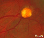
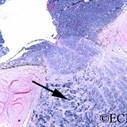
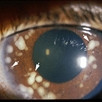
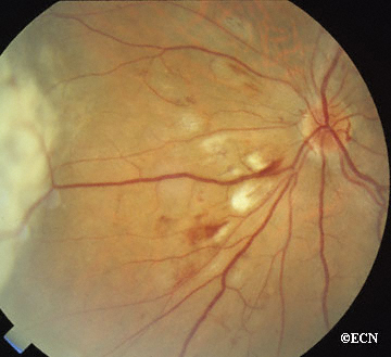
Von Hippel angioma can grow within the retina or optic nerve. They characteristically have a “feeding” retinal arteriole and a “draining” retinal vein. Bilateral involvement can be seen in up to 50% of individuals.

Von Hippel angioma are vascular tumors, not cancers and do not metastasize. Twenty percent of patients will be found to have the von Hippel Lindau Syndrome–associated with cerebellar hemangioma, pheochromocytomas, visceral cysts and renal cell carcinomas.

Symptoms

Von Hippel angioma patients either have no symptoms, or become symptomatic due to secondary retinal detachment or rarely neovascular glaucoma. The symptoms of retinal detachment are flashes of light, spots in the vision (floaters), and loss of vision. The symptoms of neovascular glaucoma are eye pain, light sensitivity, vision loss, and headache.

Diagnosis

Some patients with von Hippel angioma will have a family history of this disease. Von Hippel angioma are usually visible by dilated eye examination (ophthalmoscopy). Ultrasound can be used to measure the tumor’s size, and to evaluate for high internal reflectivity. Ophthalmoscopy typically reveals a dilated feeder artery and draining vein. An associated retinal detachment may be seen around the tumor or may be so large as to cover (obscure) an underlying von Hippel angioma.

von_h_2
Fluorescein Angiography – Note the feeder and draining vessels, as well as the bright “light-bulb” appearance.

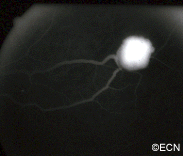
Fluorescein Angiography: Eye-care specialists perform studies of the blood vessels in the eye with a synthetic organic dye called fluorescein. The dye is injected into the arm and travels to the blood vessels inside the eye. If a tumor is in the eye, we can see specific characteristics of its circulation which can help us differentiate between it and other types of tumors. Von Hippel angiomas have a unique pattern of circulation with a feeder arteriole and a draining vein. Since the tumor extends  from the retina into the eye (vitreous humor), von Hippel angiomas exhibit intense hyperfluorescence, often compared to a “light-bulb.”

Treatments

Von Hippel angiomas can appear in both an autosomal dominant hereditary or sporadic forms. All patients should be given periodic systemic examinations including imaging studies for cerebellar hemangiomas and renal cell carcinoma. Family members should be examined with indirect ophthalmoscopy. Genetic testing is available (see related links below).

The treatment of retinal capillary hemangiomas can be a challenge to the ophthalmologist due to the presence of bilateral multiple tumors and the likelihood of new tumor formation. Despite treatment, up to 25% of cases can have permanent visual loss of acuity less to than 20/40 in one or both eyes. Various treatment modalities including observation, cryotherapy, plaque radiotherapy, and vitreoretinal surgery have been utilized.

Recent advances in the understanding of VHL protein function and tumorigenesis have led to new treatments targeting the biology of the disease, as opposed to ablative or surgical approaches. Molecules upregulated or increased in the context of a VHL mutation, such as vascular endothelial growth factor (VEGF) and platelet-derived growth factor (PDGF), have been targeted in investigational anti-angiogenic therapies, both in systemic manifestations of the disease and in ocular disease.

OBSERVATION AS TREATMENT


Observation is rarely employed due to the tendency of retinal capillary hemangiomas to progress. However, observation only might be chosen in small (Juxtapapillary hemangiomas (those next to the optic nerve disc) are particularly difficult to treat and are initially managed with observation because they can remain stable for years. As a general rule; since these tumors are not cancer and cannot metastasize; treatment should only be undertaken in case of tumor progression or a threat to visual acuity due to the adverse effect of treatment on the optic nerve and major blood vessels.

LASER PHOTOCOAGULATION

Laser photocoagulation is currently used to treat small retinal capillary hemangiomas located in the retina in eyes with clear media. When possible, we first occlude the feeder artery, then (if necessary) to surround/demarcate the posterior 180 degrees of the tumor, lastly and again if needed directly treat the tumor’s surface. Patients should be informed that multiple laser treatment sessions are typically required. Potential complications include retinal detachment, retinal and vitreous hemorrhages.

CRYOTHERAPY


Typical indications for cryotherapy are anterior retinal location of the hemangioma and massive subretinal fluid, which can reduce the laser energy uptake. Double freeze-thaw technique is employed under indirect ophthalmoscopic observation. A 15-year review found that most all hemangiomas under 3.75 mm in diameter successfully responded to cryotherapy.

ANTI-VEGF STRATEGIES


Recent studies have indicated that anti-VEGF strategies may be effective. However, no large clinical trials have been reported.

Additional Info

  1. Wong WT, Liang KJ, Hammel K, Coleman HR, Chew EY. Intravitreal ranibizumab therapy for retinal capillary hemangioblastoma related to von Hippel-Lindau disease. Ophthalmology. 2008;115:1957-1964.
  2. Annesly WJ, Leonard BC, Shields JA, Tasman WS. Fifteen year review of treated cases of retinal angiomatosis. Trans Am Acad Ophthalmol Otolaryngol. 1977;83:446-453.
  3. Madhusudan S, Deplanque G, Braybrooke JP, et al. Antiangiogenic therapy for von Hippel-Lindau disease. JAMA. 2004;291:943-944.

Collaborative Ocular Melanoma Study (COMS)

Small-sized Choroidal Melanoma Study

Small melanomas can be watched for growth prior to treatment. Should growth occur, then the patient knows the melanoma will eventually destroy the vision and increase the chance that cancer cells will spread to other parts of the body. The Collaborative Ocular Melanoma Study (COMS) was interested in how many small melanomas would grow and over what period of time. The COMS found that more than 25% of small melanomas were found to grow (within 2 years of follow-up). Since choroidal melanoma growth is the best predictor for vision loss and increased risk of metastasis, this COMS finding underscores the need to follow patients with small melanomas closely after diagnosis.

Medium-sized Choroidal Melanoma Study

The medium-sized tumor study was designed to determine if iodine-125 plaque-irradiation is better, equal, or worse than enucleation (removal of the eye) for the prevention of metastasis. In this study, half of enrolled patients were treated by enucleation and the other half underwent plaque radiation therapy. Patients were followed for evidence of recurrence and metastatic melanoma.

Collaborative Ocular Melanoma Study (COMS)
Half of enrolled patients were treated by enucleation and the other half underwent plaque radiation therapy. Patients were followed for evidence of recurrence and metastatic melanoma.

The COMS medium-tumor trial concluded that there is no significant difference between these two treatment options with respect to survival. COMS centers had followed 80% of patients for at least 5 years at the time they issued their report. Therefore, COMS found no evidence that removing the eye is a better treatment than iodine-125 plaque radiation therapy for preventing spread of choroidal melanomas.

Large-sized Choroidal Melanoma Study

Large-melanoma trial was designed to see if radiation before enucleation (removal of the eye) would prevent metastasis. The idea was to see if pre-operative irradiation would sterilize any cells that might break free during surgery. The other half of the patients did not receive radiation before their surgery.

The Large-sized Choroidal Melanoma Study concluded that patients who received 2000 rads (cGy) of external irradiation to their eye before it was removed, had an equal chance of developing metastatic disease as compared to those who were treated by enucleation (removal of the eye) alone.

Related Links


Retinoblastoma

By Paul T. Finger, MD

Description

retinoblastoma
White pupil “leukocoria” sign of retinoblastoma

Retinoblastoma is the most common intraocular cancer of childhood and affects approximately 300 children in the United States each year. More than 96% of children in North America and Europe are cured of retinoblastoma due to early detection and treatment of the affected eye. This is not true for children in countries that do not have eye cancer specialists.

Unfortunately, some children can have both eyes affected. Whenever possible, eye-cancer specialists try to save a child’s eye and preserve their vision.

Symptoms

retinoblastoma_endo-full;size$150,100.ImageHandler
An intraocular photograph of an isolated “endophytic” retinoblastoma.

Leukocoria (white pupil) and misaligned eyes (strabismus) are the most common signs of retinoblastoma. In other cases, the child may have developed neovascular glaucoma and may be in pain. Longstanding glaucoma can cause enlargement of the eye (buphthalmos). Children with neovascular glaucoma and enlargement of the eye are at greater risk for extraocular spread of their retinoblastoma.

A family history of retinoblastoma can be very important. Retinoblastoma was the first cancer to be directly associated with a genetic abnormality (Deletions or mutation of the q14 band of chromosome 13). Retinoblastoma can occur sporadically (without a family history) or it can be inherited (with a family history).

If a genetic mutation is found there is a 45-50% chance that the parents will have another child with retinoblastoma. If there is no family history and no mutation is found, the risk of having a second child with retinoblastoma is 2-5%. The average age of children first diagnosed with retinoblastoma is 18 months (typical range 0 to 36 months).

Diagnosis

More than 75% of children with retinoblastoma are first noted to have a “white-pupil” (which the doctors call leukocoria), or poorly aligned eyes (which the doctors call strabismus), or a red and painful eye (usually due to glaucoma). Other eye diseases which can cause these symptoms include congenital cataract, Toxocara canis, Coat’s disease, and persistent hypertrophic primary vitreous (PHPV). These diseases may look like retinoblastoma, but by performing an eye examination under anesthesia (EUA), specialized blood tests, digital photography, radiographic scans, and ultrasound evaluations ophthalmic oncologists can diagnose intraocular retinoblastoma in over 95% of cases. In order to be 100% correct all the time, eye-cancer specialists would have to perform a biopsy. Biopsies of intraocular retinoblastoma are avoided in order to prevent cancer cells from spreading outside of the eye.

The presence of orbital extension, uveal involvement, and optic nerve invasion are known risk factors for the development of metastatic retinoblastoma.

Treatments

Retinoblastoma treatment typically requires the cooperation of an ophthalmic oncologist, pediatric oncologist, and radiation therapist. Over the last 30 years, treatment has evolved from simple enucleation (removal of the eye), to eye-sparing radiotherapy, and more recently to chemotherapy-based multi-modality therapy (for selected cases). Intra-arterial chemotherapy (IAC) has recently been investigated to save eyes, vision and spare the child from systemic chemotherapy.

Though retinoblastoma has been cured by external beam irradiation, investigators have found that radiation may cause an increase in the risk of developing second cancers later in life.

Protocols are currently being evaluated to use chemotherapy to shrink the retinoblastoma in order to treat them with laser therapy, freezing therapy (cryotherapy), and local “plaque” radiation. Where applicable, these techniques are thought to be safer than external beam irradiation for retinoblastoma. Intra-arterial chemotherapy is a newer method of perfusing the eye with chemotherapy, used for selected cases.

Treatment of retinoblastoma often requires a team of doctors made up of ophthalmic, radiation and pediatric oncologists. These doctors should evaluate your child, discuss all the different forms of treatment, and make them available.

Related links


Low-Energy Plaque Construction

COMS-type gold seed carriers typically can be ordered in 6 sizes in 10-20 mm diameters (Trachsel DentalStudios, 1-507-288-2362).

COMS-type gold seed carriers

Rice-sized radioactive seeds are purchased and glued into the eye-plaque or seed carrier.

Rice-sized radioactive seeds

The gold of the eye-plaque will block more than 99% of the radiation to the back and sides, creating a directional source. The active surface (facing us) is sewn onto the eye beneath the base of the intraocular tumor.

The active surface of an eye plaque


Surgical Techniques for Eyelid Tumors

Description

Tumors of the eyelids may be benign cysts, inflammations (stye’s), or malignant tumors (skin cancers). The most common type of eyelid cancer is basal cell carcinoma. Most basal cell carcinomas can be removed with surgery. If left untreated, these tumors can grow around the eye and into the orbit, sinuses and brain. Other eyelid cancers include squamous cell carcinoma, sebaceous cell carcinoma, and malignant melanoma. Together, these tumors make up the remaining 10% of eyelid malignancies.

Treatment

A simple biopsy can determine if your eyelid tumor is malignant. Then, malignant tumors are completely removed and the eyelid is repaired using plastic surgery techniques. Additional cryotherapy (freezing-therapy) and radiation are sometimes required after surgery.

A Typical Work-Up for a Suspected Malignant Eyelid Tumor

  • Biopsy
  • Medical Work-up (as indicated)
  • Surgery and/or Radiotherapy of Tumor and Margins
  • Reconstruction

Surgery for Eyelid Tumors

  • General: The goal should be total removal of the cancer. This usually involves primary excision with either frozen section control or Moh’s technique.
  • Small tumors are usually removed by pentagonal wedge resection.
  • Medium-sized tumors often require reconstruction with transpositional flaps (Tenzel, Mustarde, Glabellar).
  • Large-tumor resections are typically reconstructed with Hughes, Hewes, or Cutler-Beard Techniques.

References

  1. Hughes WL. New method for rebuilding a lower lid: Report of a case. The Archives of Ophthalmology 17:1008-1017, 1937.
  2.  Hewes EH, Sullivan JH, Beard C. Lower eyelid reconstruction by tarsal transposition. American Journal of Ophthalmology 85:1164-1169, 1978.
  3. Cutler NL, Beard C. A method for partial and total upper lid reconstruction. American Journal of Ophthalmology 39:1-7, 1955.
  4. Tenzel RR, Stewart WB. Eyelid reconstruction by the semicircle flap technique. Ophthalmology 85:1164-1169, 1978.
  5. Harrington JN. Reconstruction of the medial canthus by spontaneous granulation (laissez-faire): A review. Annals Ophthalmology 14:956-960, 1982.

Related Links


Overview of the Treatment of Metastatic Melanoma

If you are newly diagnosed with a primary choroidal “intraocular” melanoma, you are likely to have no signs or symptoms of metastatic melanoma. According to a recent study utilizing total body PET/CT to stage uveal melanoma patients at diagnosis, 1% of (T1 and T2) sized tumors and 4% of (T3 and T4) size tumors were found to have their melanomas spread to other parts of their body at the time diagnosis of their eye tumor. But, up to 50% will subsequently be found to have metastasis over the following years. Be assured that many patients diagnosed and treated for choroidal melanoma will not develop metastatic melanoma.

Tumor size is the most well-verified predictor of a patient’s risk for metastatic melanoma. It makes sense that treatments that limit the tumor’s ability to enlarge will decrease the chance of metastasis. This is why most eye cancer specialists believe destroying or removing an eye cancer offers the best method to prevent future spread from that tumor.

Treatment is not thought to affect micrometastasis (too small to find) already present at the time of the eye treatment. This is why patients need periodic general medical examinations (surveys) after treatment for their intraocular melanoma.

Eighty-five percent of metastatic choroidal melanoma will be initially found in the liver. Metastases can be discovered by blood tests (liver function studies) when a patient has no symptoms. Other patients may notice abdominal fullness, discomfort and a loss of appetite. Though the liver may be the first place tumors are found, it is likely that other organs are affected. Your doctor should look for other tumor sites (e.g. subcutaneous nodules, lung, bone and brain metastasis). If a liver or skin metastasis is suspected a biopsy can be used to aspirate tumor cells for cytopathologic examination.

Since most patients start with liver tumors, therapy typically depends on the presence or absence of metastases outside of the liver, the number (size and location) of tumors within the liver, and how they affect liver function.

Treatment Options

The liver is (initially) the exclusive site of choroidal melanoma metastasis in about 40% of patients. Of those patients, most have diffuse or multi-focal tumors which cannot be removed. Treatment options depend on the size, location and rate of tumor growth.

Local Surgery: If a patient has a slow growing solitary metastasis, surgical excision may be an option. There have been no evidence-based studies that prove whether this type of surgery prolongs survival or improves the quality of life of patients. All patients who undergo surgery for a solitary liver, lung or brain metastasis have to recover from a major surgery.

Systemic Chemotherapy: When tumors are found in different parts of the body, then treatment is directed at the whole body. In these cases, your doctor may offer injection of standard intravenous chemotherapy. Unfortunately, standard chemotherapy drugs usually do not cure metastatic choroidal melanoma. There are clinical trials of new chemotherapy drugs which may be more effective.

Chemo-embolization: This treatment involves injecting a combination of chemotherapy and particles into the arteries that feed the metastatic tumors within the liver. For example, cisplatin chemotherapy and polyvinyl sponge particles are injected intra-arterially to the liver. Side effects have typically included fever, right upper quadrant abdominal pain, elevation of liver enzymes and paralysis of the intestine lasting 1 to 2 days after the procedure. It is important to understand that this is a local treatment aimed at shrinking the liver metastasis and prolonging life. It is not considered curative.

Biologic Therapy: Biologic therapy treats cancer by helping the immune system function better. The immune system is your body’s natural defense. It is a network of organs and cells distributed throughout your body. It not only defends against bacteria and viruses but also helps find and destroy cancer cells. Recent investigations focused on metastatic cutaneous melanoma have been very promising.

Observation

It is a patient’s right to choose or refuse treatment. Since many of the previously mentioned treatments can decrease a patient’s quality of life, each decision to treat must be weighed against potential side effects. You should always discuss the risk of possible side-effects and the potential benefits with your medical oncologist prior to treatment.

Related Links


Retinal Pigment Epithelium (RPE) Tumors

By Paul T. Finger, MD

Description

Adenocarcinomas and adenomas typically arise from a part of the retina called the retinal pigment epithelium or RPE. They can also occur in the ciliary body. Though all are rare, benign adenomas are much more common than RPE cancers. Less than 20 malignant adenocarcinomas of the RPE have ever been reported.  

Further, during the modern era of reporting, malignant adenocarcinomas of the RPE have never been reported to spread to other parts of the body.

In the past, almost all RPE adenocarcinomas were clinically diagnosed as atypical choroidal melanomas and the eyes were enucleated. In Dr. Finger’s review of the literature, RPE adenocarcinomas were found to be more common in females, more likely to cause intraocular inflammation (atypical for choroidal melanomas) and have responded poorly to radiation therapy.

Symptoms

Patients with adenomas of the retinal pigment epithelium typically have no symptoms. These tumors are found during routine eye examination and referred to eye tumor specialists for further evaluation. 

Diagnosis

rpe_tumors
Ultrasound of an RPE adenocarcinoma reveals a “stuck on” the choroid appearance.

Retinal adenoma and adenocarcinoma will appear black. They originate from the retina, so they are not likely to have a surrounding or overlying retinal detachment. Adenomas appear dark. “block fluorescence” on fluorescein angiography.

Ultrasound may be helpful in distinguishing adenoma and adenocarcinoma of the RPE from uveal melanomas. Consider that they tend to arise from the retina above Bruch’s membrane. Therefore, they appear to be stuck on rather than growing from the underlying choroid. Several investigators have noted a tuberous rather than collar-button or dome-shape.

Treatments

Retinal adenoma should be photographed, measured by ultrasound and followed for evidence of growth prior to consideration of treatment.

rpe_tumors_3
Histopathologic evaluation of the same tumor shows that it arises from the retina.

In the rare instance of retinal adenocarcinoma, most patients have been enucleated with a presumed diagnosis of choroidal melanoma. Should adenocarcinoma of the retinal pigment epithelium be suspected, a biopsy should be considered. Characteristics of RPE adenocarcinoma are a female patient with a dark intraocular tumor and associated with iritis and/or uveitis.


Travel Assistance Information

General Information

For starters call the National Cancer Information Center in Austin at 1-800-227-2345 and make sure you ask for a Cancer Information Specialist (CIS). The specialist must have been there a year and pass certification exams to be called a CIS.

Lodging

For lodging patients can try http://www.nahhh.org and they will provide a list of lodges/hotels/motels across the nation that offer a reduced cost place to stay.

Air Transportation

For transportation, patients can try http://www.airlifeline.org. They will try and find private pilots or commercial airlines to fly patients or provide discounts.

Financial assistance and referrals

The American Cancer Society also has a program called the “road to recovery” where volunteers will drive patients to and from appointments. When distances are great, patients may also request mileage reimbursement which may cover gas. Receipts are typically required.

Referrals for financial assistance may found at http://www.cancercare.org or call them at 1800-813-HOPE.


The Finger Classification of Radiation Retinopathy

Early radiation retinopathy
Photograph demonstrating early radiation retinopathy characterized by cotton-wool spots and intraretinal hemorrhages.

Owing to the lack of a prognosis related classification for radiation retinopathy, and the need for a common language for comparative studies, this study prompted the creation of the classification presented in the table below. There are certain preproliferative findings associated with radiation treatment of the eye.

Ophthalmoscopy is best used to view such common findings as cotton wool spots, retinal haemorrhages, ghost vessels, exudates and the less frequent retinal microaneurysms and uveal effusions. Fluorescein and indocyaninegreen angiography are typically used to define the extent of retinal ischaemia and vascular anomalies (table 5).

late radiation retinopathy
Photograph demonstrating late radiation retinopathy characterized by chorioretinal atrophy (CRA), ghost vessels (G), vascular sheathing (S), intraretinal microangiopathy (IRMA), radiation optic neuropathy (RON) and a darkened/regressed choroidal melanoma tumor (T).

When located outside the macula, stage 1 findings are consistent with excellent central vision and a good visual prognosis (mild risk). In contrast, stage 2 radiation retinopathy requires that these pathological findings are located in the macula and therefore carry a more guarded prognosis for vision (moderate risk). When the eye enters stage 3, some vision loss has probably occurred and the prognosis for return to pretreatment vision is poor (severe risk). Despite its location, the presence of retinal neovascularisation is ominous. It suggests a profound ischaemic drive and carries a worse prognosis for long term visual acuity (table 5). Vitreous haemorrhage, large areas of retinal ischaemia, and iris neovascularisation are associated with a worse prognosis for vision and globe salvage (table 5). Vitreous haemorrhage clouds our ability to use laser treatment and to monitor the progression of radiation retinopathy. Patients who present with vitreous haemorrhage often have occult neovascularisation and are at risk for ghost cell or neovascular glaucoma.

The Finger Classification of Radiation Retinopathy

The Finger Classification of Radiation Retinopathy

Table from Finger and Kurli, Br J Ophthalmol 2005;89:730–738. doi: 10.1136/bjo.2004.052159

Related Links


Enucleation: About Ocular Prosthesis Care

Q: How do I remove my prosthesis?

Ocular Prosthesis
Ocular Prosthesis

1) First, wash your hands.

2) Then you should place a towel over your lap or sink to act as a net for the prosthesis if it slips out of your hand. Should it fall it could scratch, break or get lost.

Manual Technique

3) Place one finger on the temporal (towards the ear) aspect of the lower lid on top of the cheek bone.

4) Look up.

5) Cup your other hand under your eye (to catch the prosthesis).

6) Gently press your finger in and pull the eyelid skin towards your ear (on that side).

7) The edge of your prosthesis will likely be emerging at the edge of the lower eyelid, or less likely it has fallen into your cupped hand.

8) If the prosthesis is just barely out, you can use a finger on your other hand to rotate it out of the socket.

9) Don’t be surprised if some discharge comes along with the prosthesis.

Suction Technique

1) Hard contact lens suction devices are commercially available in drug stores and vision centers.

2) These devices can be squeezed to create a vacuum that attaches the device to the front of the prosthesis.

3) Once attached, the patient can lift the bottom portion of the prosthesis out from beneath the lower lid, then slide the superior portion down towards the cheek.

Once the Prosthesis is Out

1) Commercially available sterile saline solution should be used to clean your eye socket.

2) Now you can consider cleaning the prosthesis.

Q: How do I clean my prosthesis?

1) Place the prosthetic eye into a container that can be filled with liquid as to cover the prosthesis.

2) Full or half strength hydrogen peroxide solution can be used to soak the prosthesis for 10 to 15 minutes. After soaking, remove the prosthesis from the container and rinse it with sterile saline solution.

3) Prosthesis cleaning is typically performed once or twice a week (as instructed by your eye care professionals).

4) Continuous and consistent periodic cleaning of the prosthesis will increase your comfort, decrease secretions, prevent secondary conjunctivitis and extend the life of your ocular prosthesis.

Q: How often do I need to have my prosthesis professionally cleaned or replaced?

1) You should return to your ocularist for professional cleaning and polishing 2 times each year.

2) Most patients get a new prosthesis every 3 to 5 years because even with excellent maintenance, the tissues around the prosthesis can change and the artificial eye can become scratched.

Warning

If you notice excessive discharge, swelling or irritation, you should call your eye care professional immediately.

For the more medically minded, you can go to a medical library, or order a copy of our comprehensive review of:

A Review of Enucleation
by Moshfeghi DM, Moshfeghi AA, Finger PT.

Related Links


Patient Stories

"Very well treated by Dr. Finger. He explained everything I needed to know about my issue with detail and attention, putting me at ease and giving me confidence to handle this problem for the rest of my life.”
N.N.

Read More

REQUEST AN APPOINTMENT

Go to Appointment Form

CONNECT AND SHARE