The American Joint Committee on Cancer (AJCC) Cancer Staging Manual compiles all currently available knowledge on cancer staging at various anatomic sites. In 2016, they released the 8th edition, conjunctival melanoma staging system which features 12 new staging systems, a wide range of new staging definitions, and an emphasis on the personalized-medicine approach. This staging system is used and respected by medical, ophthalmic, and radiation oncologists because it standardizes data reporting, prognosis, and selection of the best treatment for conjunctival melanoma. Nonetheless, a collaborative multicenter international registry was organized by our very own Dr. Finger to evaluate the accuracy of such a staging system. In addition to the 19 co-authors listed, Dr. Puneet Jain led the analysis, writing and collaborative revision of this study. He completed an Eye Cancer Foundation-sponsored Fellowship. ECF fellowships are known to foster ophthalmic oncology training, curiosity, learning and ability to perform research!
This study, performs the first ever international multicenter study to evaluate the validity of the eighth edition of the American Joint Committee on Cancer (AJCC) Cancer Staging Manual in estimating mortality rates of metastasis from conjunctival melanoma. The 8th edition AJCC ophthalmic oncology staging systems were written by more than 50 eye cancer specialists from 18 countries.
In this study, co-investigators utilized internet-based data sharing, reviewing 288 conjunctival melanoma patient medical histories. This study included data from 10 ophthalmic oncology centers in 9 countries over 4 continents — 2 in the United States and 1 in Canada, Colombia, Argentina, France, Netherlands, United Kingdom, Sweden, and Jordan. Clinical (cT) and pathologic (pT) staging were performed according to the staging system for conjunctival melanoma in the 8th edition of the AJCC Cancer Staging Manual.
This study was able to find new insights by conducting an analysis of large numbers of rare tumors. The findings corroborate the validity of the 8th edition of the AJCC Cancer Staging Manual. However, it also found several independent factors that are associated with increased mortality, such as tumor thickness, tumor invasion, and ulceration. Ultimately, this study supported the continued use of conjunctival melanoma staging system as published in the 8th edition of the AJCC Cancer Staging Manual.
What causes conjunctival melanoma (CoM)? Because of its rarity, much about CoM is unknown. Current medicine has yet to truly pinpoint any underlying genetic factors affecting CoM. In fact, no molecular drivers have been clearly defined in association with metastasis, recurrence prognosis, cell type, or other characteristic factors of CoM. In response to this gap in literature, a large multi-center study was launched. Over a dozen eye cancer centers collaborated in order to determine biomarkers that may indicate risk for metastasis or tumor growth.
With access to a large sample of nearly 100 patients from eight different eye cancer centers around the world, the researchers behind this study sought to clearly define key biomarkers of CoM metastatic risk, and to correlate these biomarkers to clinical features and outcomes. This association of molecular indicator to clinical feature would ultimately help treatment providers identify patients who are at greater risk for metastasis, and help researchers identify possible molecular targets for therapy.
At the conclusion of this study, it was found that in deletion mutations in a gene region called “chr10” (normally consisting of tumor-suppressing genes) significantly correlated with metastasis, lymphatic invasion, and increasing tumor thickness.
This study was the first to characterize chromosomal copy number alterations (CNAs) in such depth and with such a large and well-defined sample. The result is a more clearly-defined biomarker as a CoM risk factor than there has been in previous literature. The next plan of action with this newfound information, however, would be to apply this and other relevant research in order to further develop more effective therapies and accurate prognosis.
Check out The Eye Cancer Foundation for more information on the latest eye cancer research, charitable accomplishments, and for more information on how to donate and support new research and education!
Right alongside cancers of the breast and lung, skin cancer exists as a well-known cancer afflicting U.S. Americans. The history of cancer study has lead to the identification of over 200 types of the disease, and skin cancer is the most commonly diagnosed of them, affecting more than 1 million Americans a year. Information on this type of cancer has been widely disseminated to the American public, from a fleet of dedicated websites, to news articles, and more, to the extent that most American adults realize that skin cancer can often arise from dangerous exposure to ultraviolet (UV) sunlight. But did you know that skin cancer and eye cancer, a lesser-known type of cancer, are closely linked? Indeed, skin cancer can negatively affect the eyes — take seventy-year-old John McPartland for example, who understood this well.
In 2001, McPartland, a lifetime lover of outdoor activity, noticed a freckle on his eyelid. Determined to find answers, he consulted with many doctors until finally meeting with Dr. Paul T. Finger, who diagnosed McPartland with conjunctival melanoma. A melanoma is a particular type of skin cancer; it affects nearly 70,000 people, and is found on the melanocyte cells of the skin. Melanocyte cells are responsible for the production of brown skin pigment — melanin, Because these pigment-producing cells are afflicted, melanomas commonly begin as pigmented, odd-looking freckles like McPartland’s. The conjunctiva is a delicate, clear membrane covering the inside of the eyelids and the white (sclera) of the eye. McPartland’s diagnosis was deadly.
“I just thought I should check it out and see if it is anything,” McPartland said, “and fortunately I did … As far as I’m concerned, [Dr. Finger] saved my life.”
About 2,400 patients are diagnosed with conjunctival melanoma every year, often in part due to the same UV light that causes skin damage. Those who work outdoors, play sports and/or frequent beaches are most vulnerable to eye cancer. Having light blue eyes and a fair complexion increases vulnerability, due to a lack of melanin (brown pigment) production that protects us from harmful sun exposure. Those who have a family or personal history of skin cancer are also vulnerable. For these people, Dr. Finger recommends that they “should have an eye exam, and then every six months thereafter.”
“Certain drugs also increase UV toxicity” Dr. Finger additionally cautions. “Patients who take chlorothiazides, sulfonamides, tetracycline, phenothiazins, psoralens, and allopurinol should be extra cautious about sunlight.”
A 2008 Fox News article highlights Dr. Finger’s experience with McPartland, along with his advice for optimizing eye health that he continues to recommend to his patients today. While the importance of wearing hats and using sunblock to protect the skin has long been stressed to the American public, Dr. Finger says people should approach this thinking to the eyes as well. The best way one can optimize eye health on their own? Using UV-blocking sunglasses!
“Think of sunglasses as sunblock for your eyes”, Dr. Finger says. He advises that sunglasses with 100% UV protection offer optimal prevention of sun damage to the eye.
Cancer is certainly a difficult reality to endure for many people, but there are ways one can help to protect themselves against the disease. By doing something as simple as wearing UV-blocking sunglass, you can take charge of your health today.
Stay tuned for more stories at eyecancer.com by keeping our website in your bookmarks!
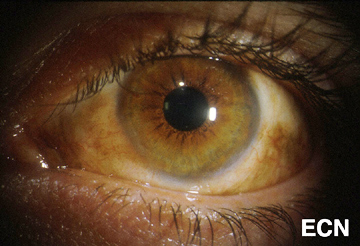
Malignant cancers can grow on the surface of the eye. They usually start from the membrane that covers most of the eye called the conjunctiva. The most common conjunctival cancers are squamous carcinoma, malignant melanoma and lymphoma.
Malignant melanoma used to be called PAM with atypia, but the newest AJCC staging system authors suggest we stop using the term PAM with atypia and call it melanoma in situ. This was because PAM with atypia doesn’t define it as a cancer. Pathologists call these specimens Tumor in situ or Tis.
Squamous carcinoma of the conjunctiva can form a nodule or diffusely spread out over the surface of the eye. Only very large squamous conjunctival cancers and those in patients who are immunosuppressed metastasize to other parts of the body. But they can invade into and around the eye, into the orbit and sinuses.
Malignant melanoma can start as a conjunctival nevus, arise as newly formed pigmentation (or variably pigmented) within the conjunctiva or onto the cornea. A simple biopsy can determine whether a pigmented conjunctival tumor is a benign nevus, primary acquired melanosis, or conjunctival melanoma.
Lymphoma can also occur in the conjunctiva. These tumors look like red or salmon-colored patches on the eye and can be the first sign of systemic lymphoma. Eye cancer specialists usually biopsy lymphoid tumors so that a pathologist can perform special immunologic and genetic studies on the tumor cells. These techniques are used to determine if the tumor is benign or malignant. Patients with lymphoid conjunctival tumors should have a complete medical check up and be examined by a hematologist-oncologist. Other “less common” conjunctival cancers are reviewed in this section.
Symptoms
Most conjunctival tumors do not cause symptoms. Patients typically seek medical attention because they notice a discoloration on the eye or extension of the tumor onto the cornea. Conjunctival tumors can also be found by an eye care specialist during a routine eye examination.
Diagnosis
Most small benign-appearing conjunctival tumors can be photographed and followed for evidence of growth prior to biopsy or treatment. If they are raised, hypervascular or extend onto the cornea a biopsy is more reasonable. At The New York Eye Cancer Center we typically obtain an office-based cytology specimen for squamous carcinoma. This lets us know the tumor is squamous and avoids a trip to the operating room. In contrast, both melanoma and lymphoma require more histopathology and special pathology analysis. Therefore, those tumors require surgical biopsy or if small, excision.
Conjunctival melanomas require special treatment. The natural history of these tumors has been characterized as presenting with multiple tumors or non-pigmented skip areas.
Therefore, it is difficult and some think impossible to define the extent of the tumor on clinical examination.
The So-Called “No Touch Technique”
The natural skip areas of pigmentation, multifocal presentation and high rates of recurrence have led some eye cancer specialists think that handling conjunctival melanoma promotes spread. They fear that tumor cells get stuck on the instruments that if reused, implant tumor on other, unaffected parts of the globe.
However, Dr. Finger disagrees with this theory and its so called “no touch technique” that requires surgeons to get new instruments after each time they touch the tumor (to prevent transplantation). However, there are no other cancers where surgeon-related transplantation has been found. More likely, these eye cancer specialists operate on one area, thinking that the pigmented portion of the tumor is the complete extent of disease. Unaware, there are other non-pigmented or small tumors that are not yet visible and later grow. Then they think they transplanted the tumor.
At The New York Eye Cancer Center, when conjunctival melanoma is suspected, Dr. Finger utilizes his specially designed cryotherapy devices “Finger-tip” cryotherapy probes” to make sure the tumor and a surround of normal appearing tissue is treated before he touches the tumor. Then the tumor can be safely removed without fear of transporting tumor cells or invaginating the edges of the wound rendering tumor too deep to treat with chemotherapy eye drops. At The New York Eye Cancer Center, topical chemotherapy eye drops are used for treatment of both squamous and melanoma cancers. These drops have been found to reduce or more commonly eliminate the need for extensive surgery.
Evaluation of the biopsy specimen should be performed by an experienced ophthalmic pathologist. If there isn’t an ophthalmic pathologist in your area, you can request that the histopathology slides be sent for second opinion.
Treatment: General Guidelines
Small tumors can be completely removed, and if they are found to be either squamous carcinoma or malignant melanoma, additional cryotherapy (freezing) is likely to improve local tumor destruction and thus prevent recurrence. Dr. Finger has developed specialized “Finger-tip” cryotherapy probes to uniformly freeze large surfaces of the eye with minimal intraocular penetration (see innovations section).
Chemotherapy Eye Drops:
Conjunctival melanoma and squamous carcinoma can be difficult to treat if they are “mulitfocal” – occur in multiple spots on the eye. In these cases, even surgical removal with freezing therapy may not control the tumor. Dr. Finger has found that “Chemotherapy eye-drops can be used for and are often better than surgery for most patients with conjunctival cancers.” Chemotherapy eye-drops treat the entire surface of the eye, are less dependent upon defining the tumors edges, and decreases the chance of scarring (symblepharon) after surgery. Researchers at The New York Eye Cancer Center recently published on treatment of “Giant Conjunctival Squamous Carcinoma’ with chemotherapy eye drops alone (no surgery).
Systemic lymphoma can usually be treated with standard chemotherapy that is also likely to cure malignant ocular lymphomas. If the eye is the only site of malignant lymphoma, low dose external beam radiation therapy is commonly employed.
"Very well treated by Dr. Finger. He explained everything I needed to know about my issue with detail and attention, putting me at ease and giving me confidence to handle this problem for the rest of my life.”
– N.N.

 With access to a large sample of nearly 100 patients from eight different eye cancer centers around the world, the researchers behind this study sought to clearly define key
 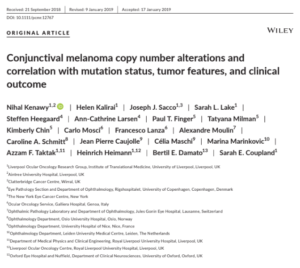 Click here for the full-text!