Malignant tumors from other parts of the body can spread in and around the eye. These tumors may never be discovered unless they affect the vision, are visible in the iris, or push the eye forward.
Symptoms
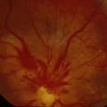
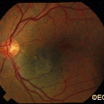
Most patients with choroidal metastasis have no symptoms, that is unless the tumor affects the central macular retina, optic nerve or the front of the eye (iris). For example, this patient’s tumor (pictured above) affected his optic nerve, caused decreased vision and floaters (spots in his vision).
Diagnosis
Most patients have a history of primary cancer (as in this case), and the tumor’s characteristics are typical of metastatic disease. This tumor was yellow-white, poorly circumscribed, less than 3 mm thick and causing congestion with vascular compromise of the optic nerve.
Treatments
As with this patient, most choroidal metastasis will resolve with external beam radiation therapy. Like many patients treated for metastatic disease, this patient received 30 Gy in 10 daily fractions. Other treatment protocols (doses) may be different, depending on the type of primary tumor.
Dr. Finger currently recommends that orbital radiation therapy for metastatic disease be performed in 180-200 cGy daily fractions. This is due to the unique sensitivity of the eye and lacrimal (tear) systems. With improvements in systemic therapy, patients are living longer and thus developing late radiation complications. Dr. Finger believes these complications will be less likely if the radiation is delivered in smaller daily fractions.
Like a raised freckle on the skin, nevi can also occur inside your eye. The most common “choroidal nevus” or eye nevus are unusual and can only be seen by an eye care specialist. Like a nevus on the skin, a choroidal nevus can grow into a malignant melanoma.
A choroidal nevus rarely requires treatment. Photography is typically used to document the size of the choroidal nevus. If the choroidal nevus has orange pigmentation, if the nevus is leaking fluid, or has a thickness of 2 mm or more it may be (or become) a malignant choroidal melanoma.
Depending on its appearance, patients with a choroidal nevus should have their eyes examined (at least) every 6 months. Only your doctor can look inside your eye to see if the choroidal nevus has changed. If the choroidal nevus has orange pigment or has thickened, it should be checked more often. If a choroidal nevus is leaking subretinal fluid, this is a particularly ominous sign. Such tumors should be followed most closely for evidence of growth or malignant transformation into a choroidal melanoma.
It is reasonable to have an eye cancer specialist check to see if your choroidal nevus looks suspicious and take baseline measurements. This examination may include the use of ultrasound, specialized photography, optical coherence tomography or an intraocular angiogram. It is a good idea for each patient to keep a picture of your choroidal nevus. This picture can be compared to future examinations to help determine if the nevus has changed or stayed the same.
Symptoms
A benign choroidal nevus (eye freckle) rarely causes symptoms. However, if a choroidal nevus leaks fluid or is associated with the growth of abnormal blood vessels (neovascularization) patients can become symptomatic. Such changes can cause a localized retinal detachment/degeneration, flashing lights and loss of vision.
A typical choroidal nevus is asymptomatic or “causes no symptoms” and found by routine dilated eye examination with ophthalmoscopy.
Diagnosis
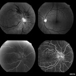
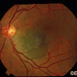
This image demonstrates how a suspicious choroidal nevus can demonstrate focal leakage on fluorescein angiography.
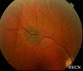
Choroidal nevus is typically a pigmented tumor of the blood vessel layer (choroid) beneath the retina. A choroidal nevus is typically gray but can be brown, yellow or variably pigmented. Your eye care professional will look to see if the choroidal nevus is raised (has thickness), orange pigment (lipofuscin), or is leaking fluid (retinal detachment). If the choroidal nevus has one or more of these findings, it is labeled a suspicious choroidal nevus that has a chance of turning into or even being a small choroidal melanoma.
A choroidal nevus can have yellow-white spots on its surface called drusen or drusenoid retinal pigment epithelial detachments (DRPED).These are signs of retinal dysfunction. The nevus may be preventing the eye from removing retinal waste products or creating microscopic leaks beneath the retina.There are no studies that show how long it takes for drusen to form on a choroidal nevus.
Treatments
A benign choroidal nevus requires no treatment and there is no way to safely remove them. Since a choroidal nevus can turn into a choroidal melanoma, it is reasonable to have it periodically observed by your eye care professional.
Additional Info
Dr. Finger believes that since skin and conjunctival melanomas have been linked to ultraviolet exposure, and since choroidal melanomas are more commonly found in patients with blue eyes, outdoor occupations, and in Australia (where there is an ozone hole); it is reasonable to wear ultraviolet (UV) blocking sunglasses. Dr. Finger says, “Think of sunglasses as sun block for your eyes.”™
"Very well treated by Dr. Finger. He explained everything I needed to know about my issue with detail and attention, putting me at ease and giving me confidence to handle this problem for the rest of my life.”
– N.N.