Dr. Paul Finger Named One of America’s Best Doctors in 2017

Dr. Paul Finger has been named one of America’s top doctors in 2017.

Castle Connolly’s top doctors are nominated by their peers through an extensive survey process of thousands of American physicians. Each year, Castle Connolly receives nearly 100,000 nominations through this process. The organization’s research team screens medical education, training, board certifications, hospital appointments, disciplinary histories, and other information to determine the best doctors in America.

This marks the 10th straight year Dr. Finger has received this honor.

“After this careful review of credentials, nominated physicians may be chosen to be listed as a regionally outstanding or a nationally outstanding Castle Connolly Top Doctor. Castle Connolly Top Doctors are highly sought after physician leaders in their communities and metropolitan areas. National Top Doctors are the top referred specialists that attract patients from wide geographical areas and across the country.”

Dr. Finger was also named among Castle Connolly’s Top Doctors for Cancer in 2017. He has earned inclusion in this list every year since its inception in 2006.

For the 15th straight year, Dr. Finger was named a Top Doctor in the New York metro area.

Inclusion among America’s top doctors reflects Dr. Finger’s commitment to his patients, and his passion for developing new and innovative eye cancer treatments.


Mother’s Persistence Leads to Retinoblastoma Diagnosis

When it comes to your children’s health, trust your instincts!

The persistence of an Oklahoma mother likely saved her daughter’s life.

When Kourtney Norton noticed her daughter McKinley was walking sideways, she knew something wasn’t right. So, Kourtney took McKinley to her pediatrician. He mistakenly thought the little girl had an eye infection. When McKinley continued exhibiting strange symptoms, Kourtney took things to the next level.

“I thought, ‘Something is going on,’” she, told KFOR.com. “She’s not really walking with her left side. She’s feeling around.”

Kourtney felt certain something more than an eye infection was causing her daughter’s symptoms. With some persistence, she got her daughter referred to an eye specialist. That led to an Oklahoma cancer center where doctors discovered a large intraocular tumor. It had already caused blindness in McKinley’s left eye and extended into her optic nerve.

Ultimately, Kourtney was diagnosed with retinoblastoma.

Doctor’s had to remove McKinley’s eye. She is now undergoing chemotherapy, but she is likely to survive, thanks to her mom’s persistence.

Retinoblastoma is the most common intraocular childhood cancer. Each year it affects approximately 300 children in the United States and 8,000 worldwide. In the USA, it is usually detected early, and the cure rate is high. But in the developing world the diagnosis is often delayed, resulting in the loss of one or both eyes, and even death. There is a hereditary link in about 40% of cases. The risk factors for sporadic retinoblastoma remain largely unknown.

Like the McKinley’s, parents need to be aware of retinoblastoma signs and symptoms. A white spot in the pupil appearing when light shines into the eye is the most common sign. This often shows up in flash photography. Other symptoms include a lazy eye, vision problems, redness in the white part of the eye, bulging eyes, changes in the color of the iris, and unequally reactive pupils.

Early screening is also important to detect cancer in its beginning stages. You should make sure your pediatrician regularly examines your child’s eyes.

Most importantly, be persistent if you think something is wrong. If you aren’t satisfied with what your doctor tells you, get a second opinion. You know your children better than anybody. Trust your instincts and be persistent. As the saying goes, it’s better to be safe than sorry.

If you are interested in helping spread awareness of retinoblastoma symptoms, click here for more information.


The Eye Cancer Foundation Hosts Successful Retinoblastoma Workshop in China

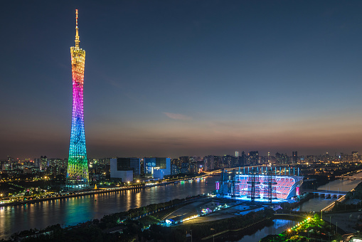
Last fall, The Eye Cancer Foundation hosted a retinoblastoma workshop in Guangzhou, China.

The primary goal was to deliver systematic training in retinoblastoma management based on the general principles of the origin and development of the disease. But the workshop went beyond practical training. It served to build strong relationships among all of the participants.

Eleven faculty members interacted enthusiastically with 19 students. The students included practicing ophthalmologists, oncologists, pathologists, and radiologists. These medical professionals already cared for retinoblastoma patients in their daily work, but they generally lacked the opportunity to discuss the science and rationale behind the treatment with others.

You can watch videos of the workshop sessions here.

The extensive effort the faculty put into their presentations was evident in the discussions, which included topics ranging from core treatment modalities, to fundamental molecular mechanisms in retinoblastoma, to social and life issues facing patients’ families. Eager students engaged in active dialogue, which even extended into breaks. The students left the workshop possessing greater depth of knowledge and new ideas for treating retinoblastoma in China.

The workshop also kicked off a long-term project. Students left with homework: all of the students filled out a table outlining their opinions of care options for a child presenting with IIRC1 Group D eye. This homework is first step in a study of informed consent for retinoblastoma families, and all participants of the workshop will be co-authors.

The Eye Cancer Foundation has a video library of the workshop talks that will be posted on the organization’s website to benefit students worldwide.

The long-term impact of the workshop is still being measured. All participating students filled out pre-workshop and post-workshop assessments to gage their knowledge of retinoblastoma. Once the videos are available in China, and students have an opportunity to review them, we will conduct additional questionnaires.

Comments from the students reveal just how valuable the workshop was.

“..these lectures in Guangzhou touched me deeply.”

“…such a wonderful workshop…very comprehensive training of retinoblastoma, covered overall content of RB from epidemiology, genetic, pathology to clinical treatment. The excellent talks and heated discussion refreshed my knowledge and will help me to improve my clinical treatment of RB.”

“So glad to have this opportunity…We learned much more updated knowledge about RB.”

“I benefited a lot from this training”

“Deeply indebted …for the comprehensive lessons in RB Workshop”

“After the training, I have a deep understanding about retinoblastoma.”

“The most important thing I learn is cooperate. We can’t fight the disease alone.”

“I learned much in this meeting and workshop. Lack of experience in diagnosis and treatment, I did not exchange with experts, but I have the chance to consult some domestic counterparts.”

ick here to watch the workshop videos.

1. Murphree AL. Intraocular retinoblastoma: the case for a new group classification. Ophthalmology clinics of North America. 2005;18:41-53.

 2. Mallipatna AC, Gallie BL, Chevez-Barrios P, Lumbroso-Le Rouic L, Chantada GL, Doz F, Brisse HJ, Munier FL, Albert DM, Catala-Mora J, Desjardins L, Suzuki S, Carroll WL, Coupland SE, Finger PT. Chapter 68: Retinoblastoma Ophthalmic Sites: Part XV  The AJCC Cancer Staging Manual, 8th edition. Editor in Chief, Mahul B. Amin, Ed. Board: Edge, SB, Byrd, DR, Compton, CC, Fritz, AG, Greene, FL, Trotti A.   New York, Springer 2017, pp. 819-831.


The Eye Cancer Foundation Retinoblastoma Workshop

GuangZhou, China November 28- 30, 2016

Purpose

  • Systematic retinoblastoma management training
  • Understand general principles of pathogenesis of retinoblastoma

Duration: 3.5 days

Trainees: 12-15 doctors

Trainers:

Dr. Junyang Zhao, Beijing Children’s Hospital, China;

Dr. Xiaoli Ma, Pediatric Oncology, Beijing Children’s Hospital, China

Dr. Bin Li, Beijing Tongren Hospital, China

Dr. Jing Zhang, Guangzhou Women and Children Medical Center, China

Dr. Brenda Gallie, Director Retinoblastoma Program, SickKids Hospital, Professor University of Toronto, Canada;

Dr. Tero Kivelä, Helsinki University Central Hospital,        University of Helsinki, Faculty of Medicine, Helsinki, Finland;

Professor David Mackey, Perth, Australia;

Professor Sandra E. Staffieri, Centre for Eye Research Australia

Dr. Francis Munier, Head, Ocular-oncology Unit, Jules Gonin Eye Hospital, Professor, Faculté de biologie et de medicine de l’UNIL, Lausanne, Switzerland;

Dr. Bhavna Chawla, Ocular Oncology Service, Rajendra Prasad Centre for Ophthalmic Sciences, All India Institute of Medical Sciences, New Delhi, India;

Dr. Vikas Khetan, Ocular Oncology Service, Senkara Nethralaya, Chennai, India;

Dr. Wantanee Dangboon, Department of Ophthalmology, Faculty of Medicine, Prince of Songkla University, Hat Yai, Songkhla, Thailand;

Dr. Pamela Astudillo, Medical Specialist III, Jose B Lingad Memorial Regional Hospital, Consultant, Angeles University Foundation Medical Center, The Philippines;

Ms. Sandra E. Staffieri, PhD Candidate, Clinical and Research Orthoptist,Centre for Eye Research and Retinoblastoma Care Co-ordinator, Department of Ophthalmology, The Royal Children’s Hospital, Melbourne, Australia.

Schedule

Day 1: Monday, November 28, 2016:

8:20-9:50           Geographic and world perspective

Dr. Junyang Zhao, Dr. Brenda Gallie, Dr. Tero Kivelä

  • (10 min) Workshop strategy and Questionnaires #1/3
  • (30 min) History of global retinoblastoma treatments: enucleation, radiotherapy, chemotherapy, focal chemotherapy modalities, etc. (Brenda Gallie)
  • (30 min) World epidemiological trends of retinoblastoma, especially in Asia-Pacific. (Tero Kivelä)
  • (20 min) History of retinoblastoma in China (Junyang Zhao)

9:50-10:10         Tea Break

10:10-11:40       2016 collaborative care and innovations

Drs. Junyang Zhao, Brenda Gallie

  • (20 min) Chinese retinoblastoma collaboration. (Junyang Zhao)
  • (30 min) 1RBW map & meeting, classification/staging. (Brenda Gallie)
  • (40 min) eCancerCareretinoblastoma (eCCrb) for patient care and research. (Brenda Gallie)

11:40-13:20       Lunch

13:20-14:50   Systemic chemotherapy

Drs. Xiaoli Ma, Dr. Bhavna Chawla

  • (30 min) Systemic chemotherapy and management. (Xiaoli Ma)
    • Mechanism of drugs, dosage, side effects.
    • (20 min) Multi-modal therapy for extra-ocular retinoblastoma. (Xiaoli Ma)
      • High-dose chemotherapy and stem cell rescue
      • Adjuvant chemo for high risk pathology
      • (20 min) Systemic chemotherapy for intra-ocular retinoblastoma. (Bhavna Chawla)
      • (20 min) Neo-adjuvant chemo for extra-ocular retinoblastoma. (Bhavna Chawla)

14:50-15:10       Tea Break

15:10-16:40 Genetic testing and counseling

Drs. David Mackie, Sandra E. Staffieri, Wantanee Dangboon,

  • (30 min) Principles of genetic knowledge and counseling. (David Mackey)
  • (20 min) Genomic forefront for the future. (David Mackey)
  • (20 min) Low vs full penetrance RB1 mutations and mosicism (Brenda Gallie)
  • (20 min) Genetic testing for retinoblastoma – the changing paradigm for clinical care. (Sandra Staffieri)
  • (20 min) Life-long surveillance for retinoblastoma and second cancers. (Wantanee Dangboon)

16:40-17:00       Tea Break

17:00-17:40       Day review and free discussion

Day 2: Tuesday, November 29, 2016:

8:20-9:50           Pathology

Drs. Xialin Xi, Brenda Gallie

  • (30 min) Pathogenesis of retinoblastoma. (Brenda Gallie)
  • (30 min) pTNM and pathology report. (Brenda Gallie)
  • (15 min) Critical observations, communication, actions. (Xialin Xi)
  • (15 min) High-risk eye pathology features & indication for adjuvant chemotherapy. (Brenda Gallie)

9:50-10:10         Tea Break

10:10-11:40       Examination staging and screening

Drs. Vikas Khetan, Tero Kivelä

  • (30 min) Presentation and diagnosis. (Vikas Khetan)
  • (30 min) 2017 TNMH vs 2005 IIRC (Vikas Khetan)
  • (15 min) Neonatal retinoblastoma. (Tero Kivelä)
  • (15 min) Trilateral retinoblastoma. (Tero Kivelä)

11:40-13:20       Lunch

13:20-14:50       Focal therapies

Drs. Francis Munier, Jing Zhang, Jungyang Zhao, Pamela Astudillo

  • (15 min) Intravitreal and Intra-arterial chemotherapy for retinoblastoma. (Francis Munier)
  • (15 min) Laser/cryo, plaque. (Francis Munier)
  • (20 min) Technique, dos and don’ts of selective intra-artery chemo. (Jing Zhang, presented by Drummond Xia)
  • (20 min) Enucleation, Myoconjunctival technique, Prosthetic conformers and eyes. (Pamela Astudillo)
  • (20 min) Intraocular tumor resection. (Junyang Zhao)

14:50-15:10       Tea Break

15:10-16:40       Clinic and Basic Research Opportunities

Dr. Tero Kivelä

  • (30 min) World surveys and analysis of outcomes (Tero Kivelä)
  • (30 min) Standardization of care (Tero Kivelä)
  • (30 min) Basic statistical principles for clinical reports (Tero Kivelä)
  • (15 min) Role of eCCRB (Brenda Gallie)

16:40-17:00       Tea Break

17:00-17:40       Day review and free discussion

Day 3: Wednesday, November 30, 2016:

8:20-9:50         Collaboration

Drs. Junyang Zhao, Jing Zhang, Xiaolin Xia, Francis Munier, Tero Kivelä, Brenda Gallie, Vikas Khetan, Bhavna Chawla, David Mackie

  • National China
  • National India
  • EuRBG
  • 1RBW

9:50-10:10         Tea Break

10:10-10:30 Genetic consultant and explanation of Impact/Geenseq reports. (Marc Zhao)

10:30-11:30     Knowledge Summary and Review

All participants

  • Questionaire #2/3

12:00                 Check-out


Six months after the Workshop, we request all participants to fill out Questionnaire #3/3, on line.


Don’t Forget Sunglasses: Think of them as Sunblock for Your Eyes

Memorial Day weekend is right around the corner. The holiday traditionally kicks off the summer season. That means boating out on the lake, lazy days at the beach, yard work, and long road trips.

As you get ready for all of your summer activities, you’ll probably run by the store and pick up some sunscreen and slather it on before you head out. While you’re at it, don’t forget to get some sunglasses. After all, you should “think of sunglasses as sunblock for your eyes.” tm

Wearing sunglasses isn’t just about making a fashion statement, or even comfort. They may actually serve as an important line of defense against eye cancer.

Sun can damage tissues in your eyes just like it does the skin. Of course, you can’t rub sunscreen on your eyes. That’s why you need to have (and wear) ultraviolet blocking sunglasses.

And ultraviolet blocking is key.

To protect your eyes, you need quality sunglasses that provide 100% UV protection. You want to wear sunglasses that block all UVA, UVB, and UVC rays. Many optical shops have a machine called a photometer that measures UV transmission through glasses. You want to make sure your sunglasses filter out all UV radiation or light under 400 nm in wavelength.

Ocular melanoma is the most common primary eye tumor in adults. Doctors diagnose around 2,000 new cases every year. These tumors are closely related to skin cancer. Since exposure to the sun is a known risk factor for skin, eyelids and conjunctival cancers, it follows that UV rays from the sun may also increase the risk of developing this form of eye cancer. There is circumstantial evidence linking exposure to sunlight and ocular melanoma. This form of eye cancer is more common in patients with blue eyes, arc-welders and those who work in outdoor occupations.

So don’t take a chance. Make sure you have sunblock for your eyes – a quality pair of sunglasses.


What to Expect: Eye Plaque Radiation Therapy (Video)

Radiation plaque therapy is the most common treatment for melanoma inside the eye.

This eye and vision sparing treatment utilizes a metallic plaque, sometime called a “radiation implant” or “radioactive source.” The doctor surgically implants the plaque on the wall of the eye, covering the base of the intraocular tumor. The implant remains in place for five to seven days, delivering a highly concentrated radiation dose to the tumor. The plaque’s location on the eye means surrounding healthy tissues get relatively less radiation exposure.

Once the plaque is in place, The New York Eye Cancer Center patient spends the rest of the treatment period at home or at a hotel. After the prescribed amount of time, the patient returns to the hospital to have the plaque safely removed.

The following video explains the procedure, what to expect, and the safety measures that must be followed during the course of treatment.


New Study Highlights Promising Treatment for Patients with Iris Melanoma

A new study by Dr. Sonal Chaugule and Dr. Paul Finger has identified a promising treatment for patients diagnosed with iris melanoma. This is a first study describing regression characteristics in published literature.

Uveal melanoma is the most common primary intraocular malignancy in adults. Iris melanoma is the rarest cancer in this family, making up only 2% to 3% of cases. However, recent studies have found that biopsy-proven iris melanomas can spread outside the eye in up to 11% of cases. These findingsIris melanoma support the treatment of iris melanomas.

In the past, most patients with melanoma of the iris were treated by removal of the tumor along with the surrounding iris. Though the tumor is removed, the surgical procedure usually leaves a giant pupil with a non-functional iris sphincter and symptoms of glare.

“After part of the iris is removed, it is like having one pupil constantly dilated, even in the sun,” Dr. Finger said.

In their new study Chaugule and Dr. Finger describe patterns of tumor regression and side effects after iris-sparing treatment for iris melanoma using palladium-103 (103Pd) plaque brachytherapy. At the New York Eye Cancer Center, plaque brachytherapy has been found to be a conservative treatment modality with low local recurrence rate. Treatment with plaque radiation to sterilize the melanoma eliminates the need to open the eye, remove the tumor and make the pupil abnormally large.

Dr. Chaugule sought to examine and document the patterns of change after plaque radiation therapy for iris melanoma. The study included 50 patients with iris melanoma who underwent 103Pd plaque brachytherapy with at least 6 months of follow-up. Pre-treatment and post-treatment tumor morphology, gonioscopy and high frequency ultrasound imaging was studied and analysed.

In this study, palladium (103Pd) plaque brachytherapy of iris melanomas showed dissappearance of blood vessels within the tumor,, darkening of tumor surface, and decreased tumor thickness. With 100% local and systemic control at a mean duration of 5.2 years, the study shows this to be a safe and effective pupil sparing treatment.

You can click here to download and read the full study.


Chinese Family Takes Long Journey to Save Daughter’s Eyes

Lulu was less than 2 months old when doctors in China diagnosed her with retinoblastoma. When doctors located tumors in her eyes, they scheduled her to have her eyes removed that afternoon. But her parents, who are both blind, were determined to do everything in their power to save their daughter’s eyes.

“My husband and I both lost our vision when we were very young,” Feifei Lin said. “Having gone through this, we have to grasp her ever-so-little remaining vision.”

In order to get their daughter treatment, the family raised over $100,000 to travel to United States.

A video produced by Stat News chronicles the family’s saga.

Their journey brought them to New York, where doctors treated Lulu using intra-arterial chemotherapy. This procedure involves treading a catheter into an artery on top of the leg and into the ophthalmic artery. This allows doctors to profuse the chemotherapy medication only into the eye.

Retinoblastoma is the most common intraocular childhood cancer. It affects approximately 300 children in the United States annually, but about 2,000 children in China are diagnosed with the disease each year. Dr. Paul Finger said lack of access to adequate care in many countries turns retinoblastoma into a deadly killer.

“In the United States, less than 2% of children die from retinoblastoma, but around the world, 70% of children die from retinoblastoma. So, what’s the difference? The difference is early diagnosis and treatment.”

The Eye Cancer Foundation is working to change that by training eye cancer specialists to work in underserved countries such as China , India, Bolivia, Vietnam, Mexico, Ethiopia, Yemen, and others.

As part of these efforts, The Eye Cancer Foundation launched its 2020 Campaign. The organization plans to train 20 eye cancer specialists to work in 20 countries by 2020. This will save thousands of children’s lives.

You can become part of the cure with a one-time or recurring donation to The Eye Cancer Foundation. Click HERE to donate today.


New Treatment Option for Patients with Radiation Macular Degeneration

A recent study published in the British Journal of Ophthalmology  by Dr. Richard Kaplan, Dr. Sonal S Chaugule, and Dr. Paul Finger introduces a new treatment option for patients with radiation macular degeneration that no can no longer be controlled with maximum, standard anti-VEGF medication.

Radiation macular degeneration is the most common cause of permanent vision loss in patients treated with plaque or proton radiation for intraocular “choroidal” melanoma. Without treatment, radiation macular degeneration causes substantial vision loss in up to two-thirds of patients at 10 years after their initial radiation therapy.

Dr. Finger’s recently published 10-year study showed that intraocular injections of anti-VEGF medications (Avastin, Lucentis) were effective in preserving vision 80% of patients. However, despite maximum doses of these medications, some patients experience progression of their macular disease.

eye treatment med

The recently published study focused on patients who were not responding to maximum doses of anti-VEGF medication. They treated by adding 4 mg of periodic intraocular triamcinolone acetonide (a steroid injection) to continued maximum anti-VEGF injections.

Kaplan and colleagues noted that adding steroid injections, stabilized or improved vision in 100% of patients at 3 months, 88% at 6 months, 88% at 9 months and 75% at 12 months. A side effect of increased eye pressure after starting the steroid injections was noted in 2 patients. However, the steroid-induced glaucoma was controlled with eye drop medications in both cases. One patient had progression of cataract.

The study authors concluded that intraocular triamcinolone steroid injection is an exciting new option to for patients who no longer respond to maximum, standard anti-VEGF treatment.


2017 Eye Cancer Working Day a Huge Success

The Second Eye Cancer Working Day, hosted by The Eye Cancer Foundation, International Society of Ocular Oncology, and American Joint committee on Cancer took place on March 24th, 2017 at International Convention Center, Sydney, Australia.

The working day provided a unique opportunity for eye cancer specialists from around the world to work together, face-to-face. The goal was to help the subspecialty move forward into the mainstream of oncological care. workind day 2

The day was divided into five sections, each dealing with important, critical problems faced by the ocular oncology specialty. Each session followed the same general format, beginning with an overview presentation by the section convenors, followed by extensive interactive group discussions. These brainstorming sessions allow participants to offer suggestions for work completion and for increasing international collaborations within each subject area.

Following is an overview of the sessions.

Session 1: Comprehensive Open Access Surgical Textbook (COAST)

Conveners: Santosh G Honavar, MD; Sonal S Chaugule, MD; Carol Shields, MD; Dan Gombos, MD; Zenyel Karcioglu, MD; Paul T Finger, MD; Hardeep Mudhar, MD

Authors who coordinated various sections of this oncology surgical guide presented their work at various levels of completion. Participants offered welcomed suggestions to make each chapter both more comprehensive and better focused toward outreach to doctors in underserved areas of the world.

Session 2: Ophthalmic Radiation Side Effect Registry (RASER)

Conveners: Wolfgang Sauerwein, MD; Paul T. Finger MD; Brenda Gallie MD

Presenters discussed information relating to a grading system for ophthalmic radiation side effects. Committed participating centers were announced, and there was an outreach to include new partners. Questions were raised that helped to modify the staging systems and create data fields for this prospective registry.

Session 3: Fellowship Outreach Retinoblastoma (FOR-RB)

Convener: Ashwin Mallipatna, MD

The proposed curriculum for fellowship training in retinoblastoma management was opened for discussion. Input from participating experts from various training institutes was documented. Excellent feedback offered by participants will be used to help finalize the first curriculum for ophthalmic oncology fellowship education.

Session 4: Doctor Reported Outcome (DRO)

Conveners: Tero Kivelä, MD; Bertil Demato, MD; Ezekiel Weis, MD

Dr. Kivelä utilized an hour-long question and answer period to help guide ophthalmic oncology toward outcome reporting. Participants discussed available methods for data collection related to DRO aimed to improve quality assurance of centers worldwide. Subjects ranging from online reporting of published outcomes to prospective collection of outcome data were also discussed. Additionally, participants considered the results of an ongoing multicenter project of Patient Reported Outcomes (PRO).

Session 5: Multicenter International Registries (MIR)

Conveners: Bita Esmaeli, MD; Brenda Gallie, MD; Martine Jager, MD

New, completed, and ongling international multicenter projects were summarised. The panel highlighted accomplishments, including retrospective registry-derived answers to important clinical questions related to choroidal melanoma staging, the failure of local control, retinoblastoma staging, and ocular adnexal lymphoma. Ongoing registries were enumerated and attendees were invited to participate. These included vitreoretinal lymphoma, conjunctival melanoma, and eyelid tumors. The process and requirements for participation of new centers in the registries was also discussed. Dr. Zeynel Karcioglu called for establishment of a chemotherapy side effects registry (in consideration of the advent of IAC and the many biotherapies with ophthalmic side effects).

The day was concluded with discussion by doctors Paul T Finger, Martin Jager, Ashwin Mallipatna, Brenda Gallie, Tero Kivelä, Wolfgang Saurwein, and Bita Esmaeli relating to future courses of action. Dr. Finger strongly suggested that the WD initiatives should be part of the International Society of Ophthalmic Oncology (ISOO), noting that most cancer subspecialties have them, and that ISOO committees need be formed to move forward.

Here is a video clip of the discussions that took place in Sydney, Australia’s, Second Working Day.


Developing a Common Language for Eye Cancer Specialists

Led by its chair Dr. Paul Finger, The Ophthalmic Oncology Task Force (OOTF) is building a foundation of multicenter, international consensus guidelines to allow for better communication and patient care.

Along with the AJCC and the Union for International Cancer Control (UICC), the American Brachytherapy Society (ABS), The American Association of Physicists in Medicine (AAPM), and The Eye Cancer Foundation (ECF) have all supported or adopted the work of the OOTF. Each has contributed to important steps toward collaboration in the field of ophthalmic oncology.AJCC Cancer Staging Manual, 8th Edition

The centerpiece of these efforts is the 8th edition of the American Joint Committee on Cancer (AJCC) Cancer Staging Manual.

AJCC-UICC tumor staging allows every eye cancer specialists to describe each patient’s cancer in a standardized way. It provides descriptions of the size, shape and distribution of the primary eye tumor, as well as the involvement of lymph nodes, or its spread within the body. Using the AJCC-UICC eye cancer classification, each medical professional can communicate the exact extent of each patient’s local and systemic cancer. As a result of an intensive a world-wide effort, the eighth edition of the AJCC Cancer Staging Manual is the most clinically useful TNM-based staging system for ophthalmic oncology.

In addition, The American Association of Physicists in Medicine Task Group-129 offered standardized methods to create eye plaques, calculate their radiation doses, describe quality assurance safety guidelines, and to review ophthalmic radiation therapy.

The American Brachytherapy Society also asked Dr. Finger to lead the OOTF in creation of consensus guidelines for plaque radiation of intraocular melanoma and for the childhood cancer retinoblastoma.

These efforts have provided a framework for multicenter, international cooperation in ophthalmic oncology.

In a recent editorial published by the American Academy of Ophthalmology, Dr. Finger explains the process that led to publication of the manual, and its importance.

“By speaking a scientific language understood by our nonophthalmic colleagues around the world, AJCC-UICC staging allows ophthalmology access to the mainstream of cancer care.”

We urge you to adopt the eighth edition of the AJCC-UICC classification system in your clinic, for research and in all eye cancer publications.

You can read the full editorial here.


Patient Stories

"Very well treated by Dr. Finger. He explained everything I needed to know about my issue with detail and attention, putting me at ease and giving me confidence to handle this problem for the rest of my life.”
N.N.

Read More

REQUEST AN APPOINTMENT

Go to Appointment Form

CONNECT AND SHARE