Second Eye Cancer Working Day Overview and Booklet

The Second Eye Cancer Working Day will take place Friday, March 24, at the International Convention Centre in Sydney, Australiaworking day book, at the Cookle Bay Room 1.

The Working Day provides an opportunity for eye cancer specialists from around the world to work together, face-to-face. The day will be divided into several sections, each dealing with a critical problem facing the specialty. Sections begin with an overview presentation from the moderators, followed by an interactive group session to brainstorm, complete work, and plan for the next meeting.

The 2017 Working Day will feature five separate committees focused on evidence-based medicine, educational programs, outreach to underserved areas, and multicenter quality assurance.

Click here for details about each of these committees and the physicians leading them.

If you can’t attend the working day, or if you are attending and want to get a preview of the day’s events, you can read the Second Working Day booklet here.

Click Here to download the booklet.


The Basics of Ophthalmic Ultrasonography (Video)

Ultrasound imaging is an effective method to visualize tumors on, within, and behind the eye. Ocular ultrasound machines generate sound waves that are reflected back by tissue in its path. The sound waves are then translated (by a computer) into an image.

Dr. Paul Finger has helped pioneer the use of ophthalmic ultrasound imaging in the diagnosis and treatment of eye cancers. In the following video, he presents the basic skills needed to perform ophthalmic ultrasound imaging. This includes the history of the evolution of these techniques as well as The New York Eye Cancer Center’s preferred practice patterns for evaluation of cancers of the eye and orbit.

Dr. Finger and his associates first described the use of 3D ultrasound for measurement of choroidal melanomas to document proper radioactive plaque placement and for extrascleral tumor extension. They also pioneered the use of computerized coronal C-scan ultrasound sections to measure the orbital portion of the optic nerve, to detect optic nerve sheath meningiomas and invasion of retinoblastoma into the optic nerve. Dr. Finger and his colleagues helped establish the use of high-frequency ultrasound (UBM) imaging for anterior segment tumors (melanocytoma, melanoma, cysts), and Dr. Finger has written on the use of high-frequency ultrasound for the diagnosis of conjunctival squamous carcinoma, periocular oncocytoma, Rosai Dorfman Disease, ciliary body adenocarcinoma and eyelid tumors.

This is important because high frequency ultrasound imaging is usually the best way to detect retinoblastoma hidden behind the iris, invasion of conjunctival tumors into the eye, and for measurement of iris and ciliary body tumors.

“High frequency ultrasound imaging has enabled eye cancer specialists to accurately measure anterior – iris and ciliary body – tumors, which in turn allows for eye and vision-sparing plaque radiation therapy,”Dr. Finger said.

This video not only provides a good overview of the techniques involved in ophthalmic ultrasonography, it will also point you to other resources where you can learn more.

“You have to know a little bit about ultrasound physics to be a great ophthalmic ultrasonographer.  You have to understand why you’re seeing what you’re seeing on the screen,” Dr. Finger said.

For more information contact us here.


2017 Eye Cancer Working Day Announcement and Schedule

International Society in Ocular Oncology and The Eye Cancer Foundation will sponsor the Second Eye Cancer Working Day on the first day of the ISOO meeting, Friday, March 24, at the International Convention Centre in Sydney, Australia, at the Cookle Bay Room 1.

The Working Day provides an opportunity for eye cancer specialists from around the world to work together, face-to-face. Our goal is to help the subspecialty working daymove forward into the mainstream of oncological care.  This will require the creation of evidence-based medicine, educational programs, outreach to underserved areas, and multicenter quality assurance.

The 2017 Working Day will feature five separate committees focused on these ongoing initiatives. These include the topics of international medical evidence, retinoblastoma fellowships, quality assurance, surgical standards, and consensus guidelines.

  • MIR: Multicenter International Registries create statistically significant evidence. These registries will improve patient care and help us defend our methods of diagnosis and treatment.
  • FOR- RB: Retinoblastoma fellowship initiative to address the worldwide RB mortality.
  • DRO: Quality assurance through Doctors Reporting Outcomes. Eye cancer specialists cannot know how to improve, unless they know the outcomes of their work.
  • COAST: A Comprehensive, open-access, consensus-based surgery text.
  • RASER: A prospective ophthalmic Radiation Side Effects Registry

The FIRST Working Day was held at The Curie Institute in Paris immediately prior to the ISOO 2015, and it was a big success.

We are excited to have the SECOND Working Day integrated with the biannual ISOO meeting. If you’re an eye cancer specialist attending the conference, be sure to mark your calendars and arrive by Thursday night!

Second Eye Cancer Working Day Schedule

Time: 8:00am – 5:00pm

Room: Cookle Bay Room 1, International Convention Centre

Convenors: Paul T Finger, Santosh G Honavar

Time Project
8:00am Registration and Coffee
8:30am – 9:00am


8:30am – 8:45am

8:45am – 9:00am

Introduction


Paul T Finger

Santosh G Honavar


9:00am – 10:00am


Comprehensive Open Access Surgical Textbook (COAST)

Convenor: Santosh G Honavar


Faculty: Fairooz P Manjandavida, Carol Shields, Zeynel Karcioglu, Mandeep Sagoo, Paul T Finger, Santosh G Honavar, Hardeep Mudhar, Sonal S Chaugule


10:00am – 11:00am


Radiation Side Effect Registry (RASER)

Convenor: Wolfgang Sauerwein


Faculty: Wolfgang Sauerwein, Paul T Finger, Brenda Gallie


11:00am – 11:30am


Morning Tea


11:30am – 12:30pm


Fellowship Outreach Retinoblastoma (FOR-RB)

Convenor: Ashwin Mallipatna


Faculty: Ashwin Mallipatna, Helen Dimaras, Brenda Gallie, Guillermo Chantada, James Muecke, Nathalie Cassoux, Santosh Honavar, John Zhao, Yacoub Yousef, Peter Gabel


12:30pm – 1:30pm Lunch
 

1:30pm – 2:30pm


Doctor Reported Outcomes (DRO)

Convenor: Tero Kivelä


Faculty: Tero Kivelä and faculty


2:30pm – 3:30pm


Multicenter International Registries (MIR)

Convenor: Bita Esmaeli


Faculty: Bita Esmaeli, Brenda Gallie, Martine Jager, Zeynel Karcioglu, Yulia Gavrylyuk, Paul T Finger


3:30pm- 4:00pm Afternoon Tea
4:00pm – 5:00pm Future Directions


Faculty: Santosh G Honavar, Martine Jager, Bita Esmaeli, Tero Kivelä, Ashwin Mallipatna, Wolfgang Sauerwein, Paul T Finger


**Please note that The ISOO Working Day workshop will be using live polling. Please ensure that you bring your mobile phone so that you can be an active part of the session.


Applying Chemotherapy Eye Drops Is Easier than It Sounds (Video)

When people hear the word chemotherapy, they generally think of intravenous medication injections to treat the whole body.  Of course, there are also chemotherapy drugs that can be administered orally in pill or liquid form. But there is another option available for treating eye cancer.

In 1993, Dr. Paul Finger and coworkers first reported on the use of Mitomycin chemotherapy eye drops to treat melanoma of the conjunctiva and cornea. Then in 2008, Dr. Finger and coworkers introduced interferon eye drops for extraocular melanoma. He realized that by placing chemotherapy agents directly onto the eye using drops, a high concentration of drug goes directly to the tumor. Topical treatment also limits the possibility of systemic (whole body) side effects.

Applying chemotherapy drops may sound complicated or intimidating, but it’s really not. In fact, most people put the drops in themselves.

This short video demonstrates the simple procedure.

As you watch the video, note the position of the patient. You also want to make sure excess medication does not get on your skin.

This video provides general information about applying chemotherapy eye drop, but as with any treatment, make sure you talk with your doctor about any questions. Not all chemotherapy eye drops are the same.

For more information contact us here.


What to Expect During Ophthalmic Plaque Radiation Therapy (Video)

Undergoing eye cancer treatment can be a scary prospect. At The New York Eye Cancer Center, we are committed to giving patients resources to make the experience as comfortable and stress-free as possible.

One way to do that is providing as much information as possible before treatment even begins. The following video shows one couple’s perspective on home-care during ophthalmic plaque radiation therapy.

EyePlaquePatientExperience from Paul T Finger on Vimeo.

Plaque radiation therapy is the most widely used treatment for choroidal melanoma and delivers a highly concentrated radiation dose to the tumor. This treatment results in relatively less radiation to surrounding healthy tissues.

Rice-sized radioactive seeds are attached within a gold or steel bowl called a plaque. This is surgically implanted to the wall of the eye, covering the base of the intraocular tumor. After surgery, the patient is typically sent home for five to seven days while the low-energy radiation sterilizes the tumor.

Dr. Finger performed plaque radiation treatment on Stanley Fishman.

“When I was first diagnosed diffuse iris melanoma, it was a rather frightening and daunting experience. Fortunately, Dr. Finger was able to guide me through the process very calmly and in a low-key fashion throughout all of the subsequent treatments and recovery.”

Now you can benefit from Stanley’s experience in this video featuring him and his wife. It provides some valuable insight on what to expect during the treatment period and also offers great tips on how to manage home care during this time.


Study Indicates Link Between Pesticide Use During Pregnancy and Retinoblastoma

A study that will be published in the April 2017 American Journal of Ophthalmology suggests parental pesticide exposure before or during pregnancy may play a role in the development of retinoblastoma.

515413906_e628a425bc_zResearchers found using products to kill insects during pregnancy, and up to 1 month before, led to a 2.8-fold higher risk of nonhereditary, unilateral retinoblastoma in children.

Retinoblastoma is the most common intraocular childhood cancer. Each year it affects approximately 300 children in the United States and 8,000 worldwide. Even with cure rates of 99% in the developed world, 70% of children continue to die of retinoblastoma in less developed countries. There is a hereditary link in about 40% of cases. The risk factors for sporadic retinoblastoma remain largely unknown.

The results of this new study dovetail with a study published in 2013. Researchers found some indication of elevated retinoblastoma risk associated with paternal pesticide exposure in the workplace during the 10 years prior to conception.

The latest study focused on maternal exposure to pesticides. Researchers conducted detailed telephone interviews with the mothers of retinoblastoma patients. The study included 282 cases (186 unilateral and 96 bilateral) from the Children’s Oncology Group. Researchers analyzed retinoblastoma risk using healthy, age-matched controls.

The study found using both household pesticide products, such as Raid, and professional lawn or landscaping services, were similarly correlated with an elevated risk of retinoblastoma. Home weed killer products were also associated with higher risk, but the results were not statistically significant.

Dr. Renelle Pointdujour Lim analyzed the study.

“Analysis of combined parental exposures for both types of retinoblastoma (in one or two eyes) also suggests that the increased risk conveyed by pesticide use was similar among different types of pesticides, regardless of indoor or outdoor use, the time at which they were used during pregnancy, and whether it was the mother or father who applied them. However, these confidence intervals were also inconclusive.”

There were limits to the study. It used a relatively small sample size. There was also an issue with “recall bias,” because researchers had to rely on the memories of the subjects they interviewed. In general, “Retrospectively collected exposure data introduces the possibility of recall bias; therefore, results should be interpreted cautiously until additional studies are conducted,” researchers said.

While current research is far from conclusive, there is certainly enough evidence to raise red flags. It is reasonable to assume that parents should be careful about pesticide exposure during pregnancy, or if they are planning to get pregnant in the near future.

“I am sure pesticide use is the cause of many health problems. All of us should be more aware of what we introduce into our environment” said Dr. Paul Finger of The New York Eye Cancer Center.


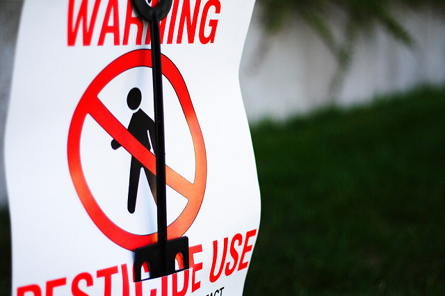
Photo by Michelle Tribe via Flickr.


A New Way to Support the Work of The Eye Cancer Foundation

There’s a new way to support the work of The Eye Cancer Foundation.

The ECF has partnered with Pledgeling, a platform that facilitates giving to nonprofits.

pledgeling5

Visitors to pledgeling.com/home can search for specific non-profit organizations or general categories they want to support. For instance, searching for “eye cancer” will bring up The Eye Cancer Foundation. They can donate directly from that page.

But Pledgeling is more than just a stop on the Internet to drop off a donation. It is a fully integrated platform allowing businesses to support charitable giving.

Pledgeling’s goal is, “Powering the revolution for good.” The organization has helped to raise over $1 million for thousands of charities since its founding in 2014.

If you own a business, you can get involved and help support important ECF projects such as our 2020 Campaign to strategically train and place eye cancer specialists in unserved regions around the world.

Pledgeling works with Evite Donations to create a platform that allows you set up giving campaigns associated with your next party or function, thereby benefitting The Eye Cancer Foundation.

“The combination of Evite’s scale with Pledgeling’s charitable giving platform has made it possible to turn any face-to-face gathering into a moment of positive social impact,” Evite CEO Victor Cho said.

Pledgeling’s mobile-first technology platform and suite of software services will allow you to seamlessly incorporate a campaign to benefit The Eye Cancer Foundation into your business. This will not only help The ECF to create a world-class resource for eye cancer patients and their families, it will also help your business. As the folks at Pledgeling put it, “brands today must do good to do well.”

You can “do good,” by helping the mission of The Eye Cancer Foundation by utilizing Pledgeling to create a campaign.

For more information on how your business can partner with the ECF through Pledgeling, click here.


Comprehensive Metastatic Screening Crucial for Ocular Melanoma Patients

Comprehensive metastatic screening in patients diagnosed with choroidal melanoma is extremely important.

Patients at the New York Eye Cancer Center have access to all screening technology commercially available. For example, PET/CT screening  was first investigated at NYECC. It is the only method that surveys the entire body for metastatic spread. In addition to an initial PET/CT scan, Dr. Paul Finger also customizes the use of subsequent MRI, CT, and ultrasound surveillance screening for his patients.

PETCTofMetastaticCancer

Unfortunately, many patients around only have access to the most basic metastatic screening. Many health services can only provide ultrasound screening. In some countries, there is no screening at all.

The American College of Radiology ranks  both abdominal contrast enhanced MRI and CT as methods superior to ultrasound for detecting metastasis to the liver. Sadly, health care systems must weigh systemic costs against individual patient care. In contrast, patients just want the best diagnostic technologies and treatments available. At the New York Eye Cancer, patient-centered care means each individual patient’s needs come first. It’s all about excellence.

Pioneered by Dr. Finger at The New York Eye Cancer Center, ophthalmic total body 18 FDG PET/CT imaging combines two technologies to create a comprehensive screening process. In simple terms, CT imaging reveals the body’s anatomy (form). Positron emission tomography (PET) uses an acceptable amount of a radioactive sugar to reveal where tissues are active (function). A complex computer program puts the form and function images together to create a complete, anatomic, and metabolic 3D reconstruction of the patients body. PET/CT allows doctors to measure tumor size and shape, show its location and effect on normal anatomic structures, as well as its metabolically activity. By overlapping the metabolic and anatomic images, for the first time PET/CT puts form and function on the same diagnostic page.

While the liver is the most common place for choroidal melanoma metastasis, a study group led by Dr. Finger found that metastases can be found outside the liver (primarily bone and skin). Ultrasound screening focuses only on the liver misses these types of metastasis.

There are many factors that indicate increased risk for metastasis in choroidal melanoma patients. Based on these risk factors, individualized diagnosis, and the patients needs, Dr. Finger determines how to approach metastatic screening for each of his patients. Once the plan is in place, he has the best tools in the world at his disposal.


The Eye Cancer Foundation Celebrates World Cancer Day

Saturday Feb. 4 marks World Cancer Day.

The theme of this year’s event is “We Can. I can.” WCD_LOGO_4C

Cancer affects people in different ways, but everybody – together and as individuals – can take action to reduce the global burden of cancer.

The ‘We can. I can.’ campaign reinforces positive and action-oriented messaging about cancer. It also encourages the engagement of organizations and individuals in raising awareness and taking action against the disease.

On World Cancer Day, The Eye Cancer Foundation will join with groups and individuals around the world committed to fighting the scourge of cancer.

World Cancer Day is an important event to spread the word and raise cancer awareness around the globe. But more importantly, it’s a springboard for action.

In the coming year, The ECF will continue pushing forward with important projects related to diagnosing, treating, and ultimately curing eye cancer.

Retinoblastoma is a life and vision-threatening eye cancer that afflicts 10,000 young children every year. Our 2020 Campaign has committed to strategically train and place eye cancer specialists in underserved regions around the world. Our immediate goal is to save 1,000 young lives by the year 2020, but with the help of our generous donors, the sky is the limit.

This year, The ECF will host the Second Working Day on the first day of the International Society of Ocular Oncology meeting March 24 in Sydney, Australia. The Working Day will bring together eye cancer specialists from around the world to continue work on multicenter, international collaboration in ocular oncology.

Another important ECF initiative is the Eye Cancer Bioinformatics Grid (BIG). The mission of the BIG is to provide an infrastructure for sharing eye cancer data, results, and research. It is the first multicenter, international cooperative program focused on improving eye cancer research, diagnosis and patient care. Bio-informatics offers the chance for eye cancer specialists to work together and pool resources. As Eye Cancer Foundation Chairman Dr. Paul Finger says, “There is power in numbers.”

At The ECF, we are proud to do our part in the broader battle against cancer, and to join together with others around the world to celebrate World Cancer Day. We can. I can make a difference in the fight against cancer.

WCD2017_WeCanICan


Knowing the Signs of Eye Cancer

Early diagnosis is one of the keys to successful treatment of eye cancer.

optometrist-91751_640

The New York Eye Cancer Center has some of the most advanced diagnostic tools and techniques available. These include high frequency and hi-resolution ultrasound imaging for tumors on, in, and behind the eye. We use high definition digital photography, and OCT imaging. In December 2015, NYECC became the first clinic in the United States to use the state-of-art OCT2 imaging machine in a clinical setting.

But all the technology in the world won’t help if patients never come in for an examination.

Eyelid, conjunctival, and corneal tumors are the easiest for patients to recognize. However intraocular and orbital tumors behind the eye are more difficult. Most patients with choroidal melanoma have no symptoms at all. These tumors are found by routine eye examinations, which highlight the need for everyone to periodically see their general ophthalmologist.

When symptoms do occur, it is crucial to know the signs and symptoms of eye cancers.

  1. Flashes of light.
  2. Squiggly lines, or floating objects.
  3. Dark spot(s) on the iris.
  4. Dark areas on the conjunctiva – the white of the eye.
  5. Bulging or swelling of the eye or eyelid.
  6. Pain in or around the eye.
  7. Loss of eyelashes
  8. Changes in the shape or size of the pupil.
  9. White pupil.
  10. Misdirection of the eye.
  11. Excessive tearing or blood in the tears.

In children, a white spot can appear in the pupil known as“leukocoria.” This is best seen when light shines into the eye and is a common sign of retinoblastoma. This often shows up in flash photography.

Of course, these symptoms don’t necessarily mean you have eye cancer, but if you notice any of them, you should visit your eye doctor as soon as possible.

Remember that eye cancer often develops with no symptoms at all. This is why annual eye exams are crucial.

By knowing the signs and symptoms, getting regular eye exams, and acting quickly if any abnormalities appear, you can drastically increase your chances of catching eye cancer in its early stages.

Early, accurate diagnosis and prompt treatment can save both your vision and your life.

In the meantime, Dr. Finger says, “Think of Sunglasses as Sunblock for your Eyes.” Tm


Patient Stories

"Very well treated by Dr. Finger. He explained everything I needed to know about my issue with detail and attention, putting me at ease and giving me confidence to handle this problem for the rest of my life.”
N.N.

Read More

REQUEST AN APPOINTMENT

Go to Appointment Form

CONNECT AND SHARE