Asking These Seven Questions Will Help You Choose the Best Doctor

It makes a difference where you are treated. Knowing the right questions to ask will help you decide.

7 Questions You Should Ask Your Doctor

Click to Read Now

We have developed a list of 7 key questions that will help you evaluate prospective physicians. Getting answers to these questions will help you be sure the doctor you chose has the experience necessary to provide the most advanced treatments. The answers will also give insight into where you (the patient) rank within the physician’s list of priorities.

When facing the uncertainty that comes with a diagnosis of eye cancer, you want a physician committed to spending all the time necessary to help you understand your condition and concerns. Discussing these 7 questions with your prospective doctors will help you determine their level of commitment. In addition, the answers will give you a peek inside their practice. You will come away with a much better sense of the doctor’s familiarity with the latest diagnostic and treatment options, how the staff views their relationship with their patients, and whether you’re likely to get caught up in bureaucratic red tape.

At The New York Eye Cancer Center, we are committed to providing patient centered care. We will happily answer these seven questions, along with any others you may have.

Click Here to see the seven questions along with more information on why they are important.


Eye Cancer Foundation Fellowship Alum Making a Difference in India

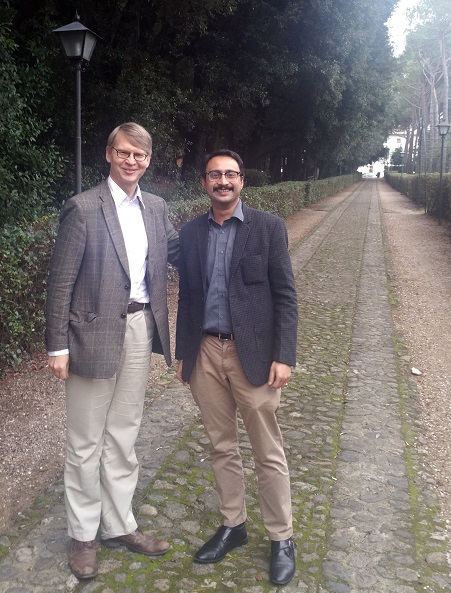
A doctor who recently completed fellowship training supported by The Eye Cancer Foundation (ECF) is providing eye cancer treatment in an area of India currently lacking any trained specialists in ocular oncology.

Dr. Bikramjit Pal worked under Dr. Tero Kivelä at the Helsinki University Eye Hospital during his six-month fellowship from October 2015 through March 2016. In addition to mentoring from Dr. Kivelä, Dr. Pal learned from a wide range of highly trained medical professionals at the hospital. He actively participated in daily patient workups in the Ocular Oncology Service and observed and assisted in the treatment of eye cancer patients.kivelapalimage2

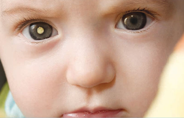
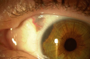
During his training, Dr. Pal was exposed to a wide range of procedures and treatments, including outpatient and intraoperative transpupillary thermotherapy, ruthenium and iodine plaque brachytherapy, conjunctival surgery, and intravitreal chemotherapy. He was involved in the treatment of conjunctival carcinoma, conjunctival melanoma, ocular adnexal and intraocular lymphoma, uveal melanoma, and retinoblastoma. He also had the opportunity to participate in one-on-one teaching in the latest diagnostic techniques related to eye cancer under the supervision of senior consultants.

In addition to his practical training, Dr. Pal also participated in research studies during his fellowship. He took part in an ocular oncology journal club and undertook three scientific projects, including one case report and two observational case series. Dr. Kivelä praised Dr. Pals work during his fellowship training:

“Dr Pal has shown keen interest in his studies, an excellent working knowledge of fundamentals of ophthalmology, and he has been a most avid observer.”

The Eye Cancer Foundation offers fellowships to doctors from unserved and underserved countries, providing specialized training in the treatment of retinoblastoma and other eye cancers. Once they complete their training, ECF fellows commit to return home to create eye cancer treatment programs.

Dr. Pal plans to begin oncology services at a tertiary eye care facility in Kolkata. This center serves a huge population, not only in eastern India, but also in the neighboring countries of Nepal and Bangladesh. Dr. Pal said he was eager to apply his training in his home country.

“In India, we deal with retinoblastoma so often; hence my aim is to start with a fully equipped clinic which will provide services to patients having intraocular tumors.”

With Dr. Kivelä’s ECF-sponsored fellowship training, Dr. Pal will be able to provide high-quality care to eye cancer patients who would otherwise have little to no access to treatment. This translates to saved vision and lives. Dr. Kivelä will continue to mentor Dr. Pal, providing long-distance consultative support and will soon visit Dr. Pal’s center.

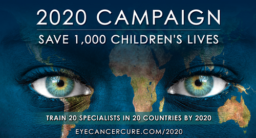
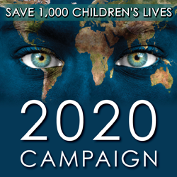
Through the recently launched 2020 Campaign, The Eye Cancer Foundation plans to multiply this success story across the world. With your help, we can train 20 eye cancer specialists to work in 20 countries by 2020.

Our immediate goal with the 2020 Campaign is to save the lives of 1,000 children by 2020. But that’s only the beginning. Our ability to train doctors and supply them with the equipment they need to properly diagnose and treat retinoblastoma is only limited by the generosity of our donors.

You can become part of the cure with a one-time or recurring donation to The Eye Cancer Foundation. Click HERE to donate today.

For more information on The Eye Cancer Foundation Fellowships, including how to apply, click HERE.


The 2020 Campaign: Train 20 Specialists in 20 Countries to Treat Childhood Eye Cancer

Every year, retinoblastoma afflicts some 10,000 children around the world. This eye cancer has a 98% cure rate with proper diagnosis and treatment, but many children in less developed countries lose their vision and even their lives because of lack of access to proper care.

Through the recently launched 2020 Campaign, The Eye Cancer Foundation plans to change that. With your help, we can train 20 eye cancer specialists to work in 20 countries by 2020. This will save thousands of children’s lives.

ecf-2020-homepage

Retinoblastoma is the most common eye cancer in children. It typically develops before the age of three. The incidence rate is somewhat higher in developing countries, where most of these children eventually succumb to metastatic retinoblastoma. In contrast, in developed countries like the US where there is access to specialized treatment, most patients survive to lead normal lives.

Screening, early diagnosis, and prompt treatment will save lives. In countries without eye cancer specialists, children with retinoblastoma typically go undiagnosed and untreated until it’s too late. Many less developed countries don’t even have a single doctor specializing in eye cancer treatment, and local general ophthalmologists are not familiar with modern eye cancer medicine. With a relatively small investment, we can provide doctors with specialized training in retinoblastoma treatment and strategically place them in underserved regions around the world.

Earlier this fall, The Eye Cancer Foundation launched a partnership with the International Council of Ophthalmology to support and promote eye cancer fellowship education for doctors from unserved and underserved countries. This partnership will greatly expand the ECF fellowship program already in place.

But we still need your help!

Through the 2020 Campaign our immediate goal is to save the lives of 1,000 children by 2020. But that’s only the beginning. Our ability to train doctors and supply them with the equipment they need to properly diagnose and treat retinoblastoma is only limited by the generosity of our donors.

You can become part of the cure with a one-time or recurring donation to The Eye Cancer Foundation. Click HERE to donate today.

Note: You are welcome to use the graphics in this post to promote the 2020 Campaign on social media or your own website. Please link to: https://www.eyecancercure.com/2020/


ecf-help-child

ecf-2020-sidebar


Unlocked Genetic Process Could Lead to New Retinoblastoma Treatment

Researchers have identified the critical role of a cancer-promoting gene in the growth and survival of retinoblastoma cells. This discovery could lead to new treatment options.

Retinoblastoma is the most common intraocular childhood cancer. It affects approximately 300 children in the United States each year and more than 8,000 worldwide. When detected early, the cure rate is high, but if the diagnosis is delayed, it can result in the loss of an eye and even death.

rb_retina_scan

Researchers at the Saban Research Institute of Children’s Hospital Los Angeles found that a protein known as MDM2 plays a critical role in the expression of another protein, MYCN, that is required for the growth and survival of retinoblastoma cells.

In previous studies, David Cobrinik, MD, PhD discovered that retinoblastoma forms from cone cells in the retina. The recent study, published in the October 17 online edition of the journal Oncogene, looked at characteristics that make these cells prone to retinoblastoma when the RB1 gene is inactivated, allowing for unchecked cell growth.

“An important way in which cone cells differ from other retinal cell types is their high expression of MDM2 and MYCN,” said first author Donglai Qi, PhD. “We have shown crosstalk between these two oncoproteins in which MDM2 promotes MYCN expression in retinoblastoma cells.”

An oncoprotein is encoded by a cancer promoting gene (oncogene). In tumor cells, oncoproteins are often mutated and expressed at high levels. Normally, MYCN provides instructions for the production of a protein that plays an important role in the formation of tissues and organs during embryonic development. But when mutated, the MYCN oncogene has the potential to cause normal cells to become cancerous.

Researchers say in addition to MCYN’s presence in retinoblastoma, it is also elevated in 20 to 25 percent of neuroblastoma tumors, and correlates with advanced disease and poor prognosis. It also plays a role in other childhood cancers.

Researchers say treatments directed at MYCN could be effective, but targeting this oncoprotein is problematic because it is difficult to block. Knowing the link between MDM2 and MYCN, researchers can now look for ways to target the mechanism through which MDM2 regulates MYCN expression.

“This identification of a critical and unexpected node in the retinoblastoma signaling circuitry could theoretically lead to pharmacologic targeting, Cobrinik said.


Easing the Way into Life After Eye Loss

Despite the many vision-saving eye cancer treatments available, sometimes it becomes necessary to remove an eye. For patients facing enucleation surgery, the prospect of losing an eye is scary. Fortunately, most patients can see with their remaining eye and will most likely be able to do all the things they used to do before, including driving a car. With modern eye prosthesis, the loss of the eye is hardly noticeable.

For patients facing enucleation surgery, or those who have already experienced the loss of an eye, the website LostEye.com serves as a fantastic resource, offering both practical information and encouragement.

Jay Adkisson created and edits LostEye.com. He had his eye surgically removed after doctors diagnosed him with choroidal melanoma. Because the tumor was so close to his optic nerve, Jay opted for enucleation. Just three years later, he earned his FAA pilot license. Jay’s testimonial on the LostEye.com home page offers encouragement for anybody facing the loss of an eye.

“Believe it or not, five days later I was back at work at my law practice, and now the fact that I only have one eye is barely an inconvenience, and not even something I notice unless I think about it. My driving isn’t any different (I give myself a little more space for the cars ahead of me), and my life isn’t any different. I haven’t even noticed any significant loss of depth perception. In other words, life has returned to pretty much what it was before I went in for the surgery.”

Jay says his experience led him to create LostEye.com as a way to help other patients.

“I know, and I mean really know – personally – how stressful losing an eye can be. Thus, I have created this completely free, noncommercial website for the information and support of those who have recently lost an eye, or who may lose an eye due to surgery, disease, etc.”

LostEye.com features a wide range of information for those facing enucleation surgery, as well as those who have already lost an eye. There is an entire page devoted to what patients can expect in the days leading up to surgery and during their recovery. You will find information on how to protect the remaining good eye and how to cope with the fear of losing an eye. There are also pages devoted to driving with one eye, prosthetic eyes, and a discussion on depth perception.

But it’s not just the information available that makes LostEye.com such a good resource. It also hosts an online community through a robust discussion forum. Here, patients facing the loss of an eye can connect with people who have already gone through the experience. They can ask questions, receive encouragement and exchange information. The opportunity to connect with others who have walked the same path helps patients realize they aren’t alone.

LostEye.com provides both information and encouragement and is a resource anybody dealing with the loss of an eye will find valuable.


Syrian Doctor Grateful for ECF Fellowship Training

Ata Rajeh, MD recently completed fellowship training in ocular oncology under Yacoub Yousef, MD of Jordan. The training was supported by The Eye Cancer Foundation (ECF).

ECF fellowships are available to candidates from unserved or underserved countries. Once the doctors complete their six months of training, they agree to return to their home country to start or participate in eye cancer treatment for the unserved.

rajeh-2

Rajeh is from the war-torn country of Syria. His fellowship ran from April to October 2016 at the King Hussein Cancer Center in Amman, Jordan. Dr. Yacoub Yousef and his team supervised Rajeh’s training. Rajeh had the opportunity to learn from a wide range of highly trained medical professionals, including retina specialists, pediatric ophthalmologists, oculoplastic surgeons, pediatric oncologists, radiotherapists, and low vision aid specialists.

Eye Cancer Foundation fellowships offer doctors specialized training in the treatment of retinoblastoma they could never receive in their home country. Retinoblastoma is the most common eye cancer in children and affects approximately 8,200 children each year. The incidence rate is somewhat higher in developing countries, where most of the children eventually succumb to metastatic retinoblastoma. In contrast, there exists a better than 96% survival rate in developed countries like the USA. Early diagnosis and treatment is key to saving retinoblastoma patients’ lives and their sight.

During his six months at King Hussein Cancer Center, Rajeh was exposed to full-range, comprehensive management of more than 100 retinoblastoma patients rajeh-1
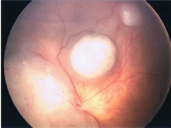
who were diagnosed at different stages in the disease. This allowed him to observe a wide variety of treatment regimens, including systemic chemotherapy, intraocular chemotherapy, intra-arterial systemic chemotherapy, focal consolidation therapies (e.g. laser and “freezing” cryotherapy), radioactive plaque therapy, external beam radiation therapy, and when necessary – removal of cancer-filled eyes.

In addition to his interaction with patients, Rajeh also accomplished research alongside Dr. Yousef. His primary project related to the management of conjunctival squamous cell carcinoma in Jordan.

“I am very excited about the results of our research,” Rajeh said. “I am the first author, and this manuscript will soon be submitted to a peer reviewed journal.”

Rajeh received invaluable training among the millions of displaced Syrians currently in Jordon. He hopes to open the first Retinoblastoma Center in Syria, and he expresses his deep gratitude to The Eye Cancer Foundation for the opportunity.

“I am going to work on building my experience in the field of ocular oncology, in the retinoblastoma field in specific,” he said in a recent letter to The Eye Cancer Foundation Board. “When I get home, I will work to establish a service that doesn’t exist in my home country. The Eye Cancer Foundation Fellowship King Hussein Cancer Center will help me a lot in gaining my aim.”

For more information on The Eye Cancer Foundation Fellowships, including how to apply, click HERE.


The International Council of Ophthalmology Highlights Fellowship Partnership with ECF

The Eye Cancer Foundation is proud to be joining forces with the International Council of Ophthalmology supporting and promoting eye cancer fellowship education for doctors from unserved and underserved countries.

double-logo

Since launching its ICO fellowship program in 2001, more than 920 Fellows have completed a three month subspecialty training and 28 ICO Fellows have completed their one year training.

This partnership between the ICO and the Eye Cancer Fellowship will expand and enhance ECF’s existing fellowship programs, focusing specifically on addressing the need to reduce world-wide mortality related to retinoblastoma by ensuring that more patients have ready access to a trained eye cancer specialist in their region.

The ICO highlighted the new in its recently released program report, Fifteen Years of ICO Fellowships. icofellowship15yearsscreenshot_1

“It is out mutual goal to decrease the high rates of world-wide mortality associated with the most common primary childhood eye cancer, retinoblastoma. Towards this goal, we agree that supplying subspecialty-trained retinoblastoma specialists to unserved and underserved countries is our priority,” The Eye Cancer Foundation Chairman Dr. Paul Finger wrote.

Together, the Eye Cancer Foundation and the ICO have set a goal of saving 1,000 children by 2020.


Drug Shows Promise in Eliminating Side-Effect of Retinoblastoma Chemotherapy

A study recently published in the Journal of Clinical Oncology showed revamping multi-drug chemotherapy for retinoblastoma to include tropotecan maintained high cure rates while preserving vision and reducing the risk of treatment related leukemia.

Retinoblastoma ranks as the most common intraocular childhood cancer. It affects approximately 300 children in the United States each year and more than 8,000 worldwide. Better than 96% of patients in North America and Europe are cured of retinoblastoma due to early detection and treatment.

eyes retino

Multi-modal chemotherapy has proved to be an effective treatment, but the widely used triple-drug therapy includes etoposide, a drug that has been associated with secondary acute myeloid leukemia in some patients.

Ten years ago, Michael Dyer, Ph.D., chair of the St. Jude Department of Developmental Neurobiology, began laboratory research seeking a safer retinoblastoma therapy. Researchers began looking at topotecan because it had shown promise in the treatment of other solid tumors.

Working with retinoblastoma tumor cells grown in the laboratory and in mice, Dyer confirmed topotecan could possibly replace etoposide in retinoblastoma therapy and determined the effective dose. The laboratory findings led to a clinical trial including 26 children diagnosed with advanced, bilateral retinoblastoma that had not spread beyond their eyes.

In the trial, triple drug chemotherapy replacing etoposide with topotecan outperformed the standard chemotherapy treatment. Of the 51 diseased eyes included in the study, 78% of those treated with therapy including topotecan were salvaged. That compares to previous reports of 30 to 60% following chemotherapy that included etoposide and often required salvage radiation therapy.

Altogether, 10 eyes had to be surgically removed, including one eye removed at diagnosis prior to chemotherapy and three removed after radiation therapy failed to stop disease progression.

“Preservation of an eye is not synonymous with preservation of vision,” first and corresponding study author Rachel Brennan, M.D. said. “But this therapy provided significant improvement in survival of eyes and useful vision in patients with advanced retinoblastoma.”

Post-treatment vision screening of survivors identified 18 patients with vision of 20/40 or better in at least one eye.

“This 10-year follow-up study shows for the first time that topotecan can be used in front-line therapy to help reduce exposure of retinoblastoma patients to etoposide,” Brennan said first. “We expected patients to be cured, but we also found more than 80 percent of patients had measurable vision.”

The results of the study seem promising. Topotecan may well provide an alternative therapy that maintains or even exceed the effectiveness of traditional multi-modal chemotherapy for retinoblastoma while eliminating one potentially deadly treatment side-effect.


Newly Developed Test Shows Promise for Assessing Metastasis Risk in Uveal Melanoma Patients

Castle Biosciences, Inc. recently launched a new test to help doctors better asses the risk of uveal melanoma metastasis.

Uveal melanoma ranks as the most common eye cancer in adults. About half of patients diagnosed with uveal melanoma develop metastatic disease, primarily in the liver.

Researchers have found gene expression levels of PRAME (Preferentially Expressed Antigen in Melanoma) correlate with an increased risk of metastasis in patients diagnosed with uveal melanoma. The newly developed gene expression profile (GEP) test measures levels of PRAME.

“Our research shows that the expression of PRAME above a specific threshold in uveal melanoma is a strong predictor of increased metastatic risk in patients with a Class 1 uveal melanoma who otherwise would be assumed to be at low risk,” University of Miami School of Medicine Dr. J. William Harbour, said. “By combining results of the gene expression profile and PRAME tests, we believe we can enhance the accuracy of metastatic risk stratification in patients with uveal melanoma.”

The test is designed to be used in conjunction with DecisionDx-UM, the company’s primary uveal melanoma test.

“DecisionDx-UM is the standard of care to identify patients whose eye tumors are likely to spread and become deadly,” President and CEO of Castle Biosciences Derek Maetzold said. “We believe the DecisionDx-PRAME test used in conjunction with the results of the DecisionDx-UM test can enable further precision of a patient’s predicted risk for metastasis and help guide physicians and patients to the most appropriate follow-up care regimens.”

In a study published last March in Clinical Cancer Research, researchers found that PRAME could distinguish between GEP Class 1 tumors that would metastasize versus those that would not. Through the study, researchers were able to determine a threshold to signify positivity in uveal melanoma tumors. Tumors that metastasized in the study were all positive for PRAME expression according to this threshold.

Castle Biosciences went on to perform technical validation of the threshold in a 958 patient sample study. In addition, researchers found patients with high metastatic risk Class 2 tumors who were positive for PRAME expression may be at higher risk of earlier metastasis compared to patients with Class 2 tumors that were PRAME negative.

Dr. Paul Finger said that while more study is necessary, the new test shows promise.

“Though we all look forward to an independent GEP and PRAME validation study, these tests are promising developments for measurement of the risk for uveal melanoma metastasis,”he said. “Early treatment and therefore early detection is key to prolonging life. Therefore this new PRAME-test should help doctors better define an individual patients risk for choroidal melanoma metastasis.”


Experimental Treatment for Ocular Melanoma Liver Metastasis Shows Promise

A new experimental treatment offers some hope for patients with ocular melanoma that has metastasized to the liver.

Ocular melanoma is the most common eye cancer in adults. About 50% of patients diagnosed with ocular melanoma go on to develop metastatic disease. In 95% of cases, the liver is the first place metastases are discovered. This is because ocular melanoma spreads through the blood-stream as opposed to the lymphatic system, making the liver susceptible to metastasis.

liver-tumor

A patient’s prognosis is generally poor once the melanoma spreads. According to the Ocular Melanoma Foundation, median survival time is between two and eight months without treatment. Standard chemotherapy tends to be ineffective, but a new experimental therapy called percutaneous hepatic perfusion (PHP) shows promise. It is now undergoing clinical trials led by the National Cancer Institute.

In PHP, doctors isolate the liver and fill it with the chemotherapy drug Melphalan. During the procedure, a catheter with a balloon on either end is inserted through several needle punctures in the skin. The balloons are then inflated, sealing off the vein above and below the liver. Once isolated, high-dose, heated chemotherapy is infused through the liver. Charcoal filters remove chemotherapy from the blood as it is returned to the body through a second catheter in the neck.

A 2010 study published in the American Journal of Clinical Oncology revealed the potential benefits of using PHP to treat both ocular and skin melanomas that have metastasized in the liver. In the study, researchers concluded the treatment significantly improved progression-free survival rates compared to the best available options.

Clinical trials have reinforced cause for optimism. In one case, Sabrina Frey, 43, recently underwent PHP treatment at Moffitt Cancer Center in Tampa, Florida. She was featured in news reports across the country and says the treatment visibly reduced her tumors.

“Some tumors are not actually even visible on my MRI’s anymore,” Sabrina said.

While PHP seems to have reduced the size of metastatic ocular melanoma tumors in the liver, it is not a cure. Researchers say the earlier the treatment is started, the better the outcome. It seems to be most successful in patients with strong liver function and a limited number of metastatic tumors.


Patient Stories

"Very well treated by Dr. Finger. He explained everything I needed to know about my issue with detail and attention, putting me at ease and giving me confidence to handle this problem for the rest of my life.”
N.N.

Read More

REQUEST AN APPOINTMENT

Go to Appointment Form

CONNECT AND SHARE