Low-Energy Plaque Construction

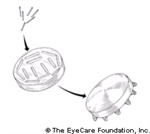
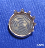
COMS-type gold seed carriers typically can be ordered in 6 sizes in 10-20 mm diameters (Trachsel DentalStudios, 1-507-288-2362).

COMS-type gold seed carriers

Rice-sized radioactive seeds are purchased and glued into the eye-plaque or seed carrier.

Rice-sized radioactive seeds

The gold of the eye-plaque will block more than 99% of the radiation to the back and sides, creating a directional source. The active surface (facing us) is sewn onto the eye beneath the base of the intraocular tumor.

The active surface of an eye plaque


Surgical Techniques for Eyelid Tumors

Description

Tumors of the eyelids may be benign cysts, inflammations (stye’s), or malignant tumors (skin cancers). The most common type of eyelid cancer is basal cell carcinoma. Most basal cell carcinomas can be removed with surgery. If left untreated, these tumors can grow around the eye and into the orbit, sinuses and brain. Other eyelid cancers include squamous cell carcinoma, sebaceous cell carcinoma, and malignant melanoma. Together, these tumors make up the remaining 10% of eyelid malignancies.

Treatment

A simple biopsy can determine if your eyelid tumor is malignant. Then, malignant tumors are completely removed and the eyelid is repaired using plastic surgery techniques. Additional cryotherapy (freezing-therapy) and radiation are sometimes required after surgery.

A Typical Work-Up for a Suspected Malignant Eyelid Tumor

  • Biopsy
  • Medical Work-up (as indicated)
  • Surgery and/or Radiotherapy of Tumor and Margins
  • Reconstruction

Surgery for Eyelid Tumors

  • General: The goal should be total removal of the cancer. This usually involves primary excision with either frozen section control or Moh’s technique.
  • Small tumors are usually removed by pentagonal wedge resection.
  • Medium-sized tumors often require reconstruction with transpositional flaps (Tenzel, Mustarde, Glabellar).
  • Large-tumor resections are typically reconstructed with Hughes, Hewes, or Cutler-Beard Techniques.

References

  1. Hughes WL. New method for rebuilding a lower lid: Report of a case. The Archives of Ophthalmology 17:1008-1017, 1937.
  2.  Hewes EH, Sullivan JH, Beard C. Lower eyelid reconstruction by tarsal transposition. American Journal of Ophthalmology 85:1164-1169, 1978.
  3. Cutler NL, Beard C. A method for partial and total upper lid reconstruction. American Journal of Ophthalmology 39:1-7, 1955.
  4. Tenzel RR, Stewart WB. Eyelid reconstruction by the semicircle flap technique. Ophthalmology 85:1164-1169, 1978.
  5. Harrington JN. Reconstruction of the medial canthus by spontaneous granulation (laissez-faire): A review. Annals Ophthalmology 14:956-960, 1982.

Related Links


Overview of the Treatment of Metastatic Melanoma

If you are newly diagnosed with a primary choroidal “intraocular” melanoma, you are likely to have no signs or symptoms of metastatic melanoma. According to a recent study utilizing total body PET/CT to stage uveal melanoma patients at diagnosis, 1% of (T1 and T2) sized tumors and 4% of (T3 and T4) size tumors were found to have their melanomas spread to other parts of their body at the time diagnosis of their eye tumor. But, up to 50% will subsequently be found to have metastasis over the following years. Be assured that many patients diagnosed and treated for choroidal melanoma will not develop metastatic melanoma.

Tumor size is the most well-verified predictor of a patient’s risk for metastatic melanoma. It makes sense that treatments that limit the tumor’s ability to enlarge will decrease the chance of metastasis. This is why most eye cancer specialists believe destroying or removing an eye cancer offers the best method to prevent future spread from that tumor.

Treatment is not thought to affect micrometastasis (too small to find) already present at the time of the eye treatment. This is why patients need periodic general medical examinations (surveys) after treatment for their intraocular melanoma.

Eighty-five percent of metastatic choroidal melanoma will be initially found in the liver. Metastases can be discovered by blood tests (liver function studies) when a patient has no symptoms. Other patients may notice abdominal fullness, discomfort and a loss of appetite. Though the liver may be the first place tumors are found, it is likely that other organs are affected. Your doctor should look for other tumor sites (e.g. subcutaneous nodules, lung, bone and brain metastasis). If a liver or skin metastasis is suspected a biopsy can be used to aspirate tumor cells for cytopathologic examination.

Since most patients start with liver tumors, therapy typically depends on the presence or absence of metastases outside of the liver, the number (size and location) of tumors within the liver, and how they affect liver function.

Treatment Options

The liver is (initially) the exclusive site of choroidal melanoma metastasis in about 40% of patients. Of those patients, most have diffuse or multi-focal tumors which cannot be removed. Treatment options depend on the size, location and rate of tumor growth.

Local Surgery: If a patient has a slow growing solitary metastasis, surgical excision may be an option. There have been no evidence-based studies that prove whether this type of surgery prolongs survival or improves the quality of life of patients. All patients who undergo surgery for a solitary liver, lung or brain metastasis have to recover from a major surgery.

Systemic Chemotherapy: When tumors are found in different parts of the body, then treatment is directed at the whole body. In these cases, your doctor may offer injection of standard intravenous chemotherapy. Unfortunately, standard chemotherapy drugs usually do not cure metastatic choroidal melanoma. There are clinical trials of new chemotherapy drugs which may be more effective.

Chemo-embolization: This treatment involves injecting a combination of chemotherapy and particles into the arteries that feed the metastatic tumors within the liver. For example, cisplatin chemotherapy and polyvinyl sponge particles are injected intra-arterially to the liver. Side effects have typically included fever, right upper quadrant abdominal pain, elevation of liver enzymes and paralysis of the intestine lasting 1 to 2 days after the procedure. It is important to understand that this is a local treatment aimed at shrinking the liver metastasis and prolonging life. It is not considered curative.

Biologic Therapy: Biologic therapy treats cancer by helping the immune system function better. The immune system is your body’s natural defense. It is a network of organs and cells distributed throughout your body. It not only defends against bacteria and viruses but also helps find and destroy cancer cells. Recent investigations focused on metastatic cutaneous melanoma have been very promising.

Observation

It is a patient’s right to choose or refuse treatment. Since many of the previously mentioned treatments can decrease a patient’s quality of life, each decision to treat must be weighed against potential side effects. You should always discuss the risk of possible side-effects and the potential benefits with your medical oncologist prior to treatment.

Related Links


Retinal Pigment Epithelium (RPE) Tumors

By Paul T. Finger, MD

Description

Adenocarcinomas and adenomas typically arise from a part of the retina called the retinal pigment epithelium or RPE. They can also occur in the ciliary body. Though all are rare, benign adenomas are much more common than RPE cancers. Less than 20 malignant adenocarcinomas of the RPE have ever been reported.  

Further, during the modern era of reporting, malignant adenocarcinomas of the RPE have never been reported to spread to other parts of the body.

In the past, almost all RPE adenocarcinomas were clinically diagnosed as atypical choroidal melanomas and the eyes were enucleated. In Dr. Finger’s review of the literature, RPE adenocarcinomas were found to be more common in females, more likely to cause intraocular inflammation (atypical for choroidal melanomas) and have responded poorly to radiation therapy.

Symptoms

Patients with adenomas of the retinal pigment epithelium typically have no symptoms. These tumors are found during routine eye examination and referred to eye tumor specialists for further evaluation. 

Diagnosis

rpe_tumors
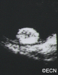
Ultrasound of an RPE adenocarcinoma reveals a “stuck on” the choroid appearance.

Retinal adenoma and adenocarcinoma will appear black. They originate from the retina, so they are not likely to have a surrounding or overlying retinal detachment. Adenomas appear dark. “block fluorescence” on fluorescein angiography.

Ultrasound may be helpful in distinguishing adenoma and adenocarcinoma of the RPE from uveal melanomas. Consider that they tend to arise from the retina above Bruch’s membrane. Therefore, they appear to be stuck on rather than growing from the underlying choroid. Several investigators have noted a tuberous rather than collar-button or dome-shape.

Treatments

Retinal adenoma should be photographed, measured by ultrasound and followed for evidence of growth prior to consideration of treatment.

rpe_tumors_3
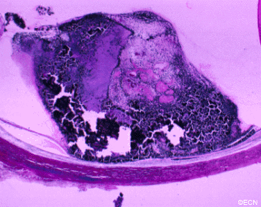
Histopathologic evaluation of the same tumor shows that it arises from the retina.

In the rare instance of retinal adenocarcinoma, most patients have been enucleated with a presumed diagnosis of choroidal melanoma. Should adenocarcinoma of the retinal pigment epithelium be suspected, a biopsy should be considered. Characteristics of RPE adenocarcinoma are a female patient with a dark intraocular tumor and associated with iritis and/or uveitis.


Travel Assistance Information

General Information

For starters call the National Cancer Information Center in Austin at 1-800-227-2345 and make sure you ask for a Cancer Information Specialist (CIS). The specialist must have been there a year and pass certification exams to be called a CIS.

Lodging

For lodging patients can try http://www.nahhh.org and they will provide a list of lodges/hotels/motels across the nation that offer a reduced cost place to stay.

Air Transportation

For transportation, patients can try http://www.airlifeline.org. They will try and find private pilots or commercial airlines to fly patients or provide discounts.

Financial assistance and referrals

The American Cancer Society also has a program called the “road to recovery” where volunteers will drive patients to and from appointments. When distances are great, patients may also request mileage reimbursement which may cover gas. Receipts are typically required.

Referrals for financial assistance may found at http://www.cancercare.org or call them at 1800-813-HOPE.


The Finger Classification of Radiation Retinopathy

Early radiation retinopathy
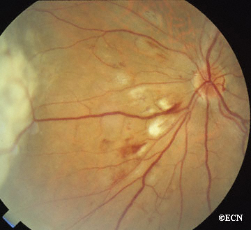
Photograph demonstrating early radiation retinopathy characterized by cotton-wool spots and intraretinal hemorrhages.

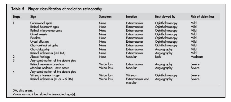
Owing to the lack of a prognosis related classification for radiation retinopathy, and the need for a common language for comparative studies, this study prompted the creation of the classification presented in the table below. There are certain preproliferative findings associated with radiation treatment of the eye.

Ophthalmoscopy is best used to view such common findings as cotton wool spots, retinal haemorrhages, ghost vessels, exudates and the less frequent retinal microaneurysms and uveal effusions. Fluorescein and indocyaninegreen angiography are typically used to define the extent of retinal ischaemia and vascular anomalies (table 5).

late radiation retinopathy
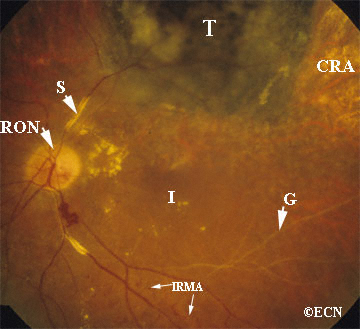
Photograph demonstrating late radiation retinopathy characterized by chorioretinal atrophy (CRA), ghost vessels (G), vascular sheathing (S), intraretinal microangiopathy (IRMA), radiation optic neuropathy (RON) and a darkened/regressed choroidal melanoma tumor (T).

When located outside the macula, stage 1 findings are consistent with excellent central vision and a good visual prognosis (mild risk). In contrast, stage 2 radiation retinopathy requires that these pathological findings are located in the macula and therefore carry a more guarded prognosis for vision (moderate risk). When the eye enters stage 3, some vision loss has probably occurred and the prognosis for return to pretreatment vision is poor (severe risk). Despite its location, the presence of retinal neovascularisation is ominous. It suggests a profound ischaemic drive and carries a worse prognosis for long term visual acuity (table 5). Vitreous haemorrhage, large areas of retinal ischaemia, and iris neovascularisation are associated with a worse prognosis for vision and globe salvage (table 5). Vitreous haemorrhage clouds our ability to use laser treatment and to monitor the progression of radiation retinopathy. Patients who present with vitreous haemorrhage often have occult neovascularisation and are at risk for ghost cell or neovascular glaucoma.

The Finger Classification of Radiation Retinopathy

The Finger Classification of Radiation Retinopathy

Table from Finger and Kurli, Br J Ophthalmol 2005;89:730–738. doi: 10.1136/bjo.2004.052159

Related Links


Enucleation: About Ocular Prosthesis Care

Q: How do I remove my prosthesis?

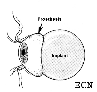
Ocular Prosthesis
Ocular Prosthesis

1) First, wash your hands.

2) Then you should place a towel over your lap or sink to act as a net for the prosthesis if it slips out of your hand. Should it fall it could scratch, break or get lost.

Manual Technique

3) Place one finger on the temporal (towards the ear) aspect of the lower lid on top of the cheek bone.

4) Look up.

5) Cup your other hand under your eye (to catch the prosthesis).

6) Gently press your finger in and pull the eyelid skin towards your ear (on that side).

7) The edge of your prosthesis will likely be emerging at the edge of the lower eyelid, or less likely it has fallen into your cupped hand.

8) If the prosthesis is just barely out, you can use a finger on your other hand to rotate it out of the socket.

9) Don’t be surprised if some discharge comes along with the prosthesis.

Suction Technique

1) Hard contact lens suction devices are commercially available in drug stores and vision centers.

2) These devices can be squeezed to create a vacuum that attaches the device to the front of the prosthesis.

3) Once attached, the patient can lift the bottom portion of the prosthesis out from beneath the lower lid, then slide the superior portion down towards the cheek.

Once the Prosthesis is Out

1) Commercially available sterile saline solution should be used to clean your eye socket.

2) Now you can consider cleaning the prosthesis.

Q: How do I clean my prosthesis?

1) Place the prosthetic eye into a container that can be filled with liquid as to cover the prosthesis.

2) Full or half strength hydrogen peroxide solution can be used to soak the prosthesis for 10 to 15 minutes. After soaking, remove the prosthesis from the container and rinse it with sterile saline solution.

3) Prosthesis cleaning is typically performed once or twice a week (as instructed by your eye care professionals).

4) Continuous and consistent periodic cleaning of the prosthesis will increase your comfort, decrease secretions, prevent secondary conjunctivitis and extend the life of your ocular prosthesis.

Q: How often do I need to have my prosthesis professionally cleaned or replaced?

1) You should return to your ocularist for professional cleaning and polishing 2 times each year.

2) Most patients get a new prosthesis every 3 to 5 years because even with excellent maintenance, the tissues around the prosthesis can change and the artificial eye can become scratched.

Warning

If you notice excessive discharge, swelling or irritation, you should call your eye care professional immediately.

For the more medically minded, you can go to a medical library, or order a copy of our comprehensive review of:

A Review of Enucleation
by Moshfeghi DM, Moshfeghi AA, Finger PT.

Related Links


Think of Sunglasses as Sunblock for Your Eyes ™

Dr. Finger advises patients on the ways in which they can avoid eye cancers, including eye cancers caused by radiation from the sun.

Ultraviolet Radiation

The sun gives off many forms of energy. Visible light from the sun helps us see the world around us. Other forms of light are not visible to the eye. These include ultraviolet (UV) light and infrared (IR) light. Even though these forms of light are not visible to the eye, they are still absorbed by the eye and the eyelids.

When all forms of light are absorbed by the body, a reaction occurs which results in heat and chemical changes. Ultraviolet light rays are particularly energetic and cause more chemical reactions (damage) in ocular tissues than visible light.

Ultraviolet (UV) Light Exposure Contributes To:

  • Eye Cancers
  • Benign Growths on the Eye
  • Corneal Burns
  • Cataract
  • Solar Retinopathy
  • Macular Degeneration

Drugs That Can Increase UV Toxicity:

  • Chlorothiazides
  • Sulfonamides
  • Tetracycline
  • Phenothiazines
  • Psoralens

If you are taking any of these drugs, care should be taken to reduce your exposure to ultraviolet light (e.g. sunlight).

Occupational Exposure to UV is Related To Sun Exposure

  • Truck Drivers
  • Mailpersons
  • Couriers
  • Pilots
  • Lifeguards
  • Farmers
  • Anglers
  • Astronauts
  • Ski Instructors
  • Park Rangers
  • Police Officers
  • Construction Workers

These occupations (among others) as well as recreational exposure can increase your risk.

Sun Block

Ultraviolet radiation is divided into UVA, UVB, and UVC. Sun block is primarily used to block UVB from burning our skin and causing cancer. SPF generally means Sun Protection Factor for UVB rays. That is SPF 8 means that if a person normally develops sunburn in 15 minutes, it will take 2 hours (8 times 15 minutes) before they burn. Some new sunblocks also stop UVA exposure, but only the opaque zinc oxide and titanium dioxide offer total protection by blocking all light.

Sunglasses

Dr. Finger says, “THINK OF SUNGLASSES AS SUNBLOCK FOR YOUR EYES.” ™

Sunglasses should block all UVA, UVB, and UVC rays. Be careful and ask for 100% UV protection. Your optical shop should have a machine that measures UV transmission through glasses called a “photometer.” The photometer should find that your sunglasses block all UV radiation or light under 400 nm in wavelength.

Sun Blocking Clothes

There are specialty clothes and hats that can be used to block Ultraviolet Radiation. For more information contact http://SunPrecautions.com.

FAQ’s

Q: What color sunglasses should I choose?

A: You may choose any color (gray, brown, green, or yellow). Some colors will affect your color vision. If you have a color vision problem gray is best (especially for driving). You can have a clear UV blocking coating on your regular glasses.

Q: What else can I do to decrease glare?

A: Polarizers and antireflective coatings can be added to your glasses to decrease glare.

Q: Do cataract implants block UV light?

A: Yes, new implants (IOL’s) contain UV blocking agents. Make sure to ask your doctor before your surgery.

Related Links


ACRONYMS: For patients treated for metastatic choroidal melanoma

  • BCNU – Carmustine
  • DTIC – Dacarbazine
  • ELND – Elective Lymph Node Dissection
  • FNAB – Fine Needle Aspiration Biopsy
  • IE or CE – Immunoembolization/Chemoembolization
  • IFN or INF – Interferon alfa-2b
  • IHP – Isolated Hepatic Perfusion
  • IL-2 – Interleukin-2
  • ILP – Isolated Limb Perfusion
  • IND – Investigational New Drug
  • Intron-A – Interferon alfa-2b
  • MRI – Magnetic Resonance Imaging
  • NED – No Evidence of Disease
  • PET – Positron Emission Tomography
  • CT – Computed Tomography
  • RFA – Radiofrequency Ablation
  • SNB or SLNB – Sentinel Node Biopsy/Sentinel Lymph Node Biopsy
  • SLN – Sentinel Lymph Node
  • TNF – Tumor Necrosis Factor
  • US – Ultrasound
  • WLE – Wide Local Excison

Do You Speak Ocular Tumor?
From The 2008 AAO Course In Atlanta

Paul T Finger, MD (Senior Instructor) Committee Chair, The Ophthalmic Oncology Task Force The American Joint Committee on Cancer- International Union Against Cancer (AJCC-UICC) Instructors: Darryl Ainbinder, MD, Sarah Coupland, MD, James C. Fleming, MD, J. William Harbour, MD, Leonard M. Holbach, MD, Zeynel Karcioglu, MD, Tero Kivelä MD, Hugh McGowan, MD, A. Linn Murphree, MD, Jack Rootman, MD, Stefan Seregard, MD, Valerie White, MD, Christian Wittekind, MD, Goupei Yu, Ph.D.

Language is defined as the human use of spoken or written words as a communication system. Sharing a common language allows us to communicate our ideas and enables progress.

With this in mind, the American Joint Committee on Cancer (AJCC), the International Union Against Cancer (UICC), and the American College of Surgeons (ACS) have come together to sponsor a committee to design a clinically useful TNM (Tumor-Node-Metastasis) based classification for ocular tumors. The AJCC classification system can be used as a universal language for those who diagnose and treat ocular tumors.

Chaired by Paul T. Finger, MD, the AJCC-UICC committee of physicians included specialists in ophthalmic pathology, ocular plastic surgery, intraocular, orbital, and adnexal tumors. Dr. Finger “Internationalized” the committee as to include members from the USA and 10 from other countries.

These included members of the UICC and others with particular expertise based on a review of the published literature. The training of committee members includes: clinical ophthalmic oncology, ophthalmic pathology, molecular biology, genetics, retinal surgery, orbital surgery and biostatistics. In a peer-review process, over 38 reviewers have been involved shaping this work.

All specialists were familiar with current methods of diagnosis and treatment. Thus, every attempt was made to create a clinically useful TNM-based classification system. TNM also means that these classifications conform to what is used throughout medicine, for other tumors and in tumor registries around the world. Clearly, the use of the AJCC-UICC TNM classification would bring ophthalmic oncology into the mainstream of cancer research.

Like Esperanto (created by Dr. L.L. Zamenhof in 1887 to function as an international language), the AJCC-UICC classification is meant to function as a universal eye cancer classification “language” system.1 Esperanto means hopeful. Clearly, we are hopeful that all scientists, clinicians and journals adopt this classification.

Why should we all adopt a single classification system for ocular tumors?

Until we all use the same classifications of ocular tumors, our studies are not directly comparable and the accuracy of meta-analysis is degraded. If we all decide to “speak ocular tumor,” we will embrace an opportunity to allow for accurate comparisons of our investigational studies.

In 1997, Dr. Finger’s review of the literature on “Radiation Therapy for Choroidal Melanoma,” pointed out that no two centers (outside the Collaborative Ocular Melanoma Study [COMS]) published using the same tumor classification.2 It was not possible to state that tumors of equivalent size and location were irradiated at each center.

Though important information was obtained by comparing multiple phase-I clinical studies, if all the investigators had used the same tumor-classification-language, it would have improved this comparative analysis. It was not until the COMS that over 40 centers used the same definitions (classifications) of choroidal melanoma size, methods to document location, and radiation dosimetry.3 In fact, the COMS is an excellent example of how a staging system can be used at multiple institutions to allow treatments to be directly comparable and additive. But, the COMS only studied choroidal melanomas. In contrast, the AJCC classification provides clinically useful definitions of tumor size, location and metastatic disease for almost all ocular cancers.4

Why is it important to compare research studies?

We all know that the wheels of progress grind exceedingly slow, but having a common ocular tumor language is an opportunity to grease that wheel. If we all use a common classification for ocular tumors, we will be able to compare treatments on equivalently sized and “staged” tumors. Such information has the potential to improve both our research and clinical decisions.

For example, it would not be reasonable to compare the use of topical mitomycin chemotherapy for multifocal conjunctival and corneal melanoma vs. another study that recruited mostly localized bulbar conjunctival tumors.

Advocates for the AJCC-UICC classification suggest that when a researcher publishes on new treatment for conjunctival melanoma, the reader should be able to discern the relative size, location, and distribution of the tumors treated. Thus, independent clinical studies can be found to be either additive or dissimilar. Our clinical and research decisions are based on ideas and studies and what we have heard in lectures or read in the literature. Clearly, these decisions are only as good as the information we acquire.

Can speaking “Ocular Tumor” affect informed consent?

Absolutely! Much of our time as clinicians is spent explaining the current knowledge and ophthalmic practice to our patients. A typical explanation of the risks and benefits includes what has been proven by statistics-based-research and what is offered as traditional practice. For example, the Reese-Ellsworth classification categorized the size, location and distribution of retinoblastoma in order to prognosticate the effect of external beam radiation therapy.5 Similarly, the AJCC-UICC tumor classifications will allow us to tell our patients that tumors of certain sizes and/or locations are more or less likely to respond to treatments and/or exhibit side effects.

How can we get everyone to employ this common language?

Unlike Esperanto, the AJCC-UICC classification should become mandatory for peer-reviewed publications. This could be done through the instructions for authors (as have other structural elements). We also request that our scientific organizations (e.g. AAO, ARVO, ISER, ASOPRS, ISOO and others) require that scientific data be submitted in the AJCC format. This may sound a bit drastic, but the editors of the major journals and officers of our organizations could make this decision to the benefit of our profession. We also request that our hospitals require that all tumors be AJCC-UICC staged prior to surgery. This would simplify the task of tumor registries all over the world. The data from tumor registries can be used to monitor both the incidence and prevalence of eye cancers.

We hope that peer reviewed journals, web sites, organizations and lay publications support this effort. If the world-wide ophthalmic community learns how to speak and publish in “Ocular Tumor,” we will better understand each other’s work and more effectively help our patients with eye cancer.

Message From the Editor
Reprinted (in part) from Ophthalmology, Vol. 110, Issue 1, 2003, pp 13-14
copyright (2003) with permission from Elsevier Science.

References

  1. Zamenhof LL. Fundamento de Esperanto, 9th ed. Marmande, France: Esperantaj Francaj Eldonoj, 1963.
  2. Finger PT. Radiation therapy for choroidal melanoma. Surv Ophthalmol 1997;42:215–32.
  3. The COMS randomized trial of iodine 125 brachytherapy for choroidal melanoma, II: characteristics of patients enrolled and not enrolled. COMS Report No. 17. The Collaborative Ocular Melanoma Study Group. Arch Ophthalmol 2001;119:951–65.
  4. Haik B, Ainbinder DJ, Finger PT, et al. Part XI: Ophthalmic sites. In: Greene FL, Page DL, Fleming ID, et al., eds. AJCC Cancer Staging Manual, 6th ed. New York: Springer, 2002:347–84.
  5. Reese AB, Ellsworth RM. The evaluation and current concept of retinoblastoma therapy. Trans Am Acad Ophthalmol Otolaryngol 1963;67:164–72.

Primary Authors:

COL. Darryl Ainbinder, MD (USA)
Sarah E Coupland, MBBS. Ph.D. (UK)
James “Chris” Fleming, MD (USA)
J. William Harbour, MD (USA)
Leonard M Holbach, MD (UICC) (Germany)
Zeynel Karcioglu, MD (USA)
Tero Kivela, MD (Finland)
Hugh McGowan, MD (Canada)
A Linn Murphree, MD (USA)
Jack Rootman, MD (Canada)
Stefan Seregard, MD (Sweden)
Valerie White, B.Sc., MD (Nfld), FRCPC (Canada)
Christian Wittekind, MD (UICC) (Germany)
Guopei Yu, Ph.D. (USA – Statistician)

Reviewers/Participants:

Daniel Albert, MD (USA)
James O. Armitage, MD (USA)
James J. Augsburger, MD (USA)
Nikolas Bechrakis, MD ( Germany)
John H. Boden, MD (USA)
Patricia Chevez-Barrios, MD (USA)
Bertil Damato, MD (UK)
Didi de Wolff-Rouendaal, MD, PhD (The Netherlands)
Laurence Desjardins, MD (France)
Ralph Eagle, MD (USA)
Deepak Edward, MD (USA)
Bita Esmaeli, MD (USA)
Brenda Gallie, MD (Canada)
Dan Gombos, MD (USA)
Jean-Daniel Grange, MD (France)
Hans Grossniklaus, MD (USA)
Barrett Haik, MD (USA)
COL John Halligan, MD (USA)
George Harocopos, MD (USA)
John Hungerford, MD (UK)
Martine Jager, MD (The Netherlands)
Emma Kujala, MD (Finland)
COL Robert A. Mazzoli, MD (USA)
Tatyana Milman, MD (USA)
Tim Murray, MD (USA)
Andrew Schachat, MD (USA)
Arun Singh, MD (USA)
Matthew Wilson, MD (USA)

Section Editor:

Frederick Greene, MD
7th Edition AJCC Cancer Staging Manual Editors
Edge SE, Byrd DR, Carducci MA, Compton CA

AJCC-UICC Administration:

Ms. Karen Pollitt
Ms. Donna Gress

Publisher

Springer, New York, USA

Chair

Paul T. Finger, MD, FACS
The AJCC-UICC Ophthalmic Oncology Task Force
Tel: 1212-832-8170

Related Links

Search Google for Do You Speak Ocular Tumor? The 11/10/2008 AAO Course in Atlanta


Patient Stories

"Very well treated by Dr. Finger. He explained everything I needed to know about my issue with detail and attention, putting me at ease and giving me confidence to handle this problem for the rest of my life.”
N.N.

Read More

REQUEST AN APPOINTMENT

Go to Appointment Form

CONNECT AND SHARE