Cavernous Hemangioma of the Orbit
By Paul T. Finger, MD
Description

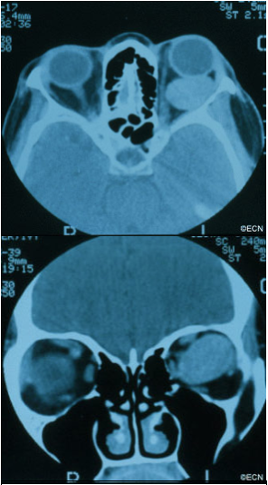
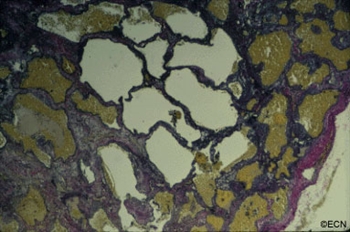
Hemangioma is a benign tumor that is found to grow within the orbit. Most commonly located behind the eye globe, it can push the eye forward causing eye-bulging doctors call proptosis.
Symptoms
Cavernous hemangioma of the orbit is most commonly seen in middle-aged women. Most are found within the muscle cone, but can be found anywhere in the orbit.
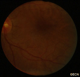
These orbital tumors can indent the back of the eye causing choroidal folds, or push on the optic nerve causing damage (atrophy).
Rarely, the tumor can push the eye so far that the cornea cannot be covered by the eye lids. In these cases, corneal exposure problems (keratitis, superficial punctate keratopathy, ulceration, even perforation) can occur.
Diagnosis
Cavernous hemangioma of the orbit is usually a slow-growing tumor. If the tumor has not damaged the eye, cavernous hemangioma can be observed for growth prior to considering intervention. Should tumor growth occur, it will be measured by eye examinations including (but not limited to) visual acuity, color vision assessment, Hertel exophthalmometry (a measure for proptosis), as well as an evaluation for double vision (strabismus), corneal exposure, retinal damage, vascular damage, and optic neuropathy.
Treatment
Treatment of orbital hemangioma is indicated when there is evidence of growth, optic nerve compression, and corneal exposure (with secondary keratitis sicca), or evidence of vision loss.
The goal of orbitotomy for choroidal hemangioma should be complete removal of the tumor. This usually involves careful dissection of the tumor to protect the tumor’s capsule (as possible). Connecting vascular feeder vessels should be identified and cauterized. A lateral orbitotomy can be required to keep the large tumor intact.


