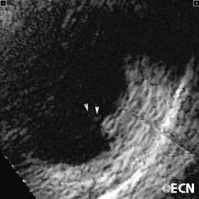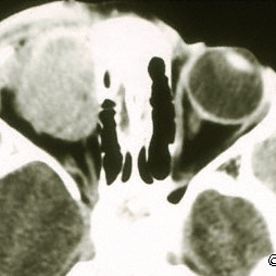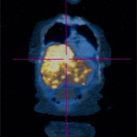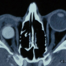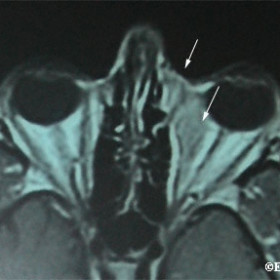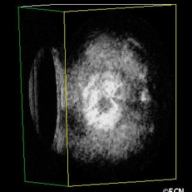
3D ultrasound
3D ultrasound demonstrates the relative position of an eye plaque utilizing a coronal section at the posterior plaque surface.
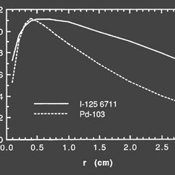
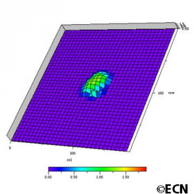
A graph of the relative dose distribution
A graph of the relative dose distribution from a single iodine-125 versus palladium-103 seed.
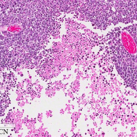
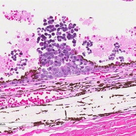
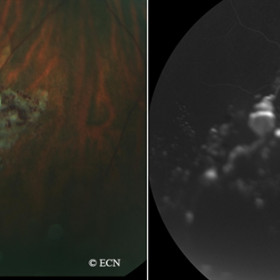
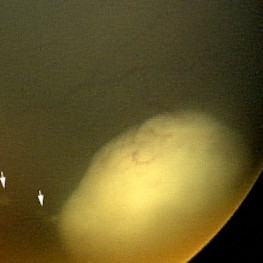
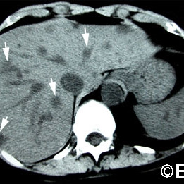
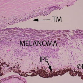
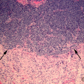
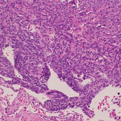
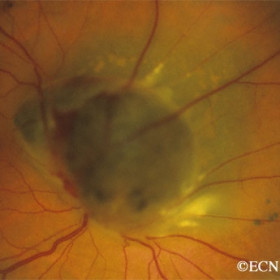
Choroidal Melanoma
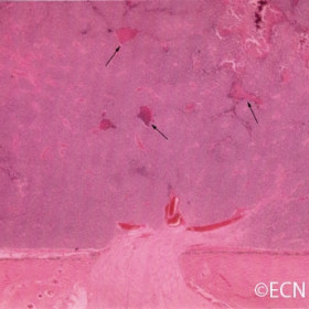
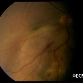
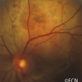
Hemorrhage (arrows) and ghost vessels are seen on the retinal surface of a juxtapapillary melanoma (4 years after treatment with palladium-103 plaque therapy). Note there is no significant radiation maculopathy.
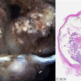
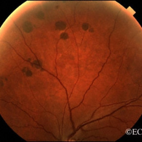
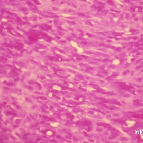
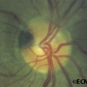
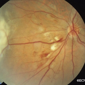
Choroidal Melanoma
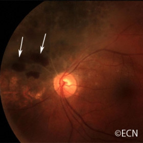
Choroidal Melanoma - Radiation retinopathy findings including, retinal hemorrhages, exudates, and ghost vessels.
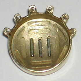
COMS gold eye plaque
COMS gold eye plaque with COMS-type silicone insert filled with iodine-125 seeds.
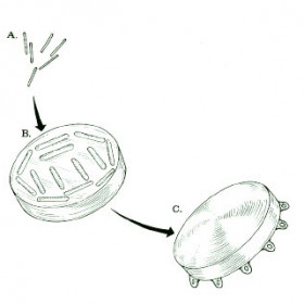
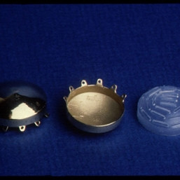
COMS plaque components
COMS plaque components - seeds, insert and gold plaque
COMS plaque
COMS plaque with palladium-103 seeds in acrylic

COMS style ophthalmic plaques
A set of COMS style ophthalmic plaques
COMS type eye plaque
COMS type eye plaque - Left shows back surface, middle reveals inner surface, right is a silicone seed holder (insert)
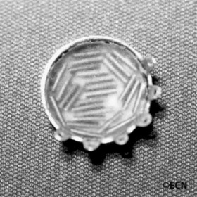
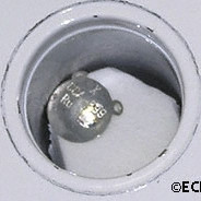
COMS type gold eye plaque
COMS type gold eye plaque with new
Seed-Guide
insert, currently holding 3 iodine-125 seeds.
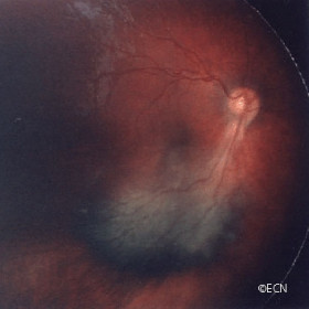
Cystoid macular edema
Cystoid macular edema can tumor or radiation therapy induced.

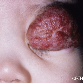
4-lights around a choroidal melanoma
Diode lights attached to an eye plaque demonstrate 4-lights around a choroidal melanoma.
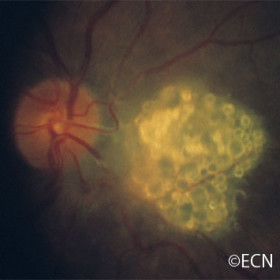
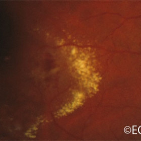
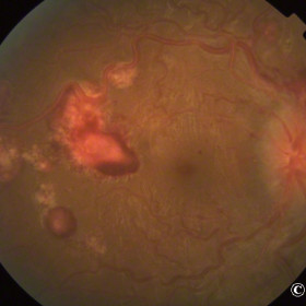
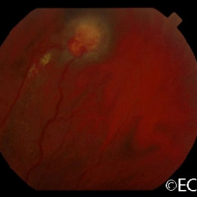
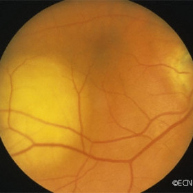
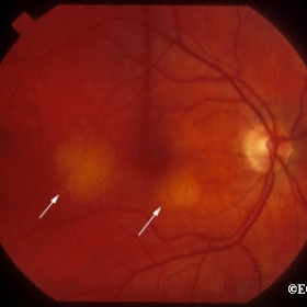
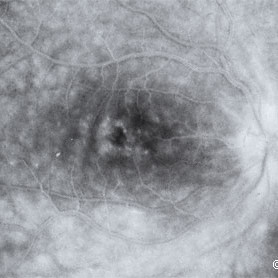
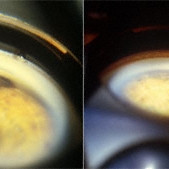
Early radiation retinopathy
Cotton wool spots and intraretinal hemorrhages are signs of early radiation retinopathy.
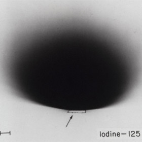
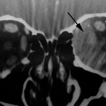
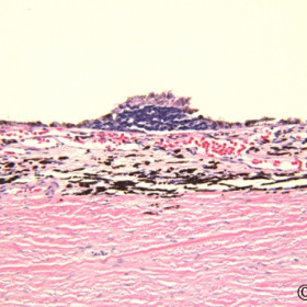
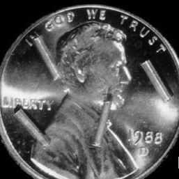
Plaque block radiation (black) on x-ray film
The gold backing and side walls of the plaque block radiation (black) on x-ray film
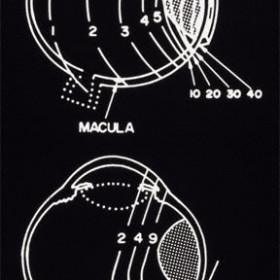
Shift in radiation fields
Graphic demonstrates the shift in radiation fields for proton treatment of anterior versus posterior choroidal melanoma
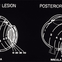
The relative radiation distribution of proton versus plaque radiation
Graphic of the relative radiation distribution of proton versus plaque radiation for an anterior T2:medium sized choroidal melanoma
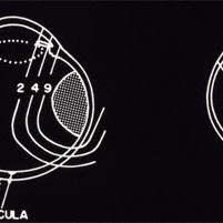
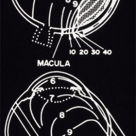
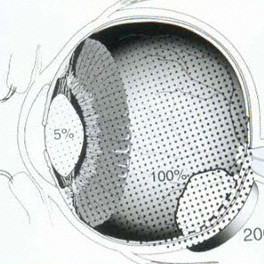
How plaque radiation distribution shifts
Graphic demonstrates how plaque radiation distribution shifts along with tumor position (note an increased dose to the macula in treatment of a posterior choroidal melanoma).
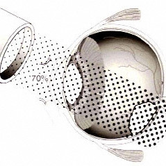
The relative radiation distribution of proton versus plaque radiation
Graphic of the relative radiation distribution of proton versus plaque radiation for a posterior T2:medium sized choroidal melanoma
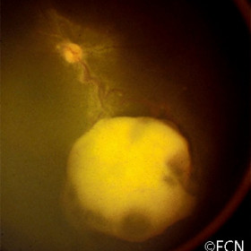
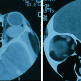
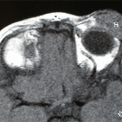
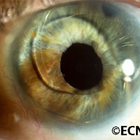
Iridociliary melanoma
Iridociliary melanoma before (left) and after (right) palladium-103 plaque radiation therapy
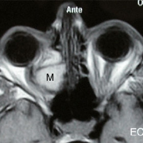
Iridociliary melanoma
Iridociliary melanoma 10-years after palladium-103 plaque radiation therapy
Iridociliary melanoma
Iridociliary melanoma 7.5 years after palladium-103 plaque radiation therapy
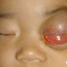
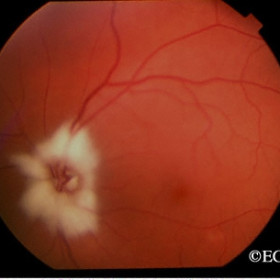
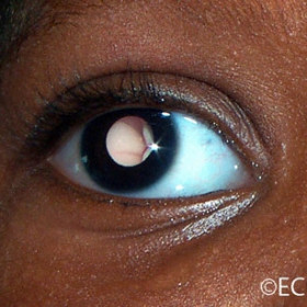
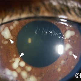
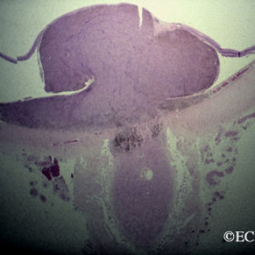
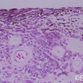
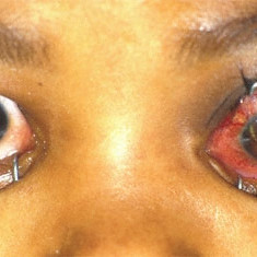
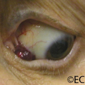
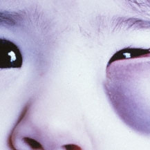
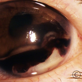
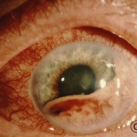
Iris Bombe
Iris Bombe- Neovascularization, posterior synechiae and secondary glaucoma can occur after irradiation for choroidal melanoma.
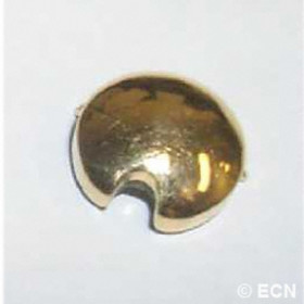
Notched COMS plaque
Notched COMS plaque with side wall (Posterior Aspect)
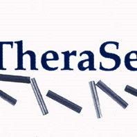
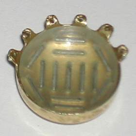
Palladium-103 seeds
Palladium-103 seeds - "Theraseed" - Promotional image
Palladium-103 seeds
Palladium-103 seeds - Note they are the same size as iodine-125 seeds but have flattened ends.
Plaque radiation therapy
Graphic of plaque radiation therapy in treatment of a T2:medium-sized posterior choroidal melanoma
Proton beam radiation therapy
Proton beam radiation therapy Eyelash loss, iris neovascularization, dry eye and cataract as can be seen after proton beam radiation therapy.
Proton radiation therapy
Graphic of proton radiation therapy in treatment of a T2:medium-sized posterior choroidal melanoma
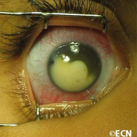
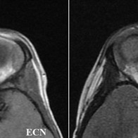
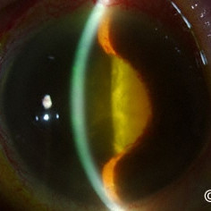
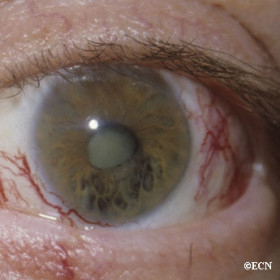
Radiation associated posterior subcapsular (PSC) cataract
Radiation associated posterior subcapsular (PSC) cataract - This unilateral cataract was seeen after iodine-125 plaque radiation therapy.
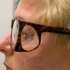
Radiation blocking glasses
Radiation blocking glasses (lateral view) can be used instead of a lead patch during low energy plaque radiation therapy.
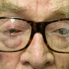
Radiation blocking glasses
Radiation blocking glasses containing lead glass can be worn during plaque therapy.
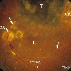
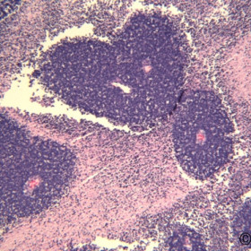
Radiation effects
Radiation effects on the posterior segment of the eye include: T= regressed tumor, CRA = choroiretinal atrophy, G= ghost vessels, S= sheathed vessels, IRMA=intraretinal microangiopathy, RON=radiation optic neuropathy
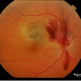
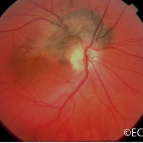
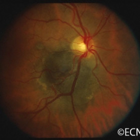
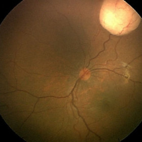
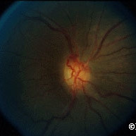
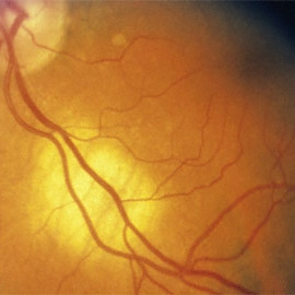
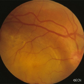
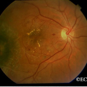
Radiation optic neuropathy
Radiation optic neuropathy - Fundus photograph demonstrates hemorrhagic variant.
Ruthenium-106 plaque in a lead pig
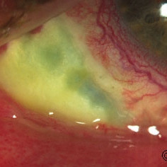
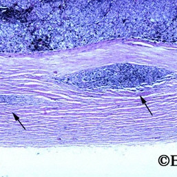
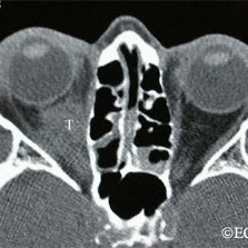
Scleral necrosis seen after ruthenium
Scleral necrosis seen after ruthenium -106 plaque radiation therapy for an anterior choroidal melanoma- An unusual complication.

USC-style gold plaque
A USC-style gold plaque with grooves to aid (standardize) seed placement.
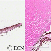
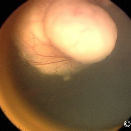
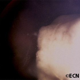
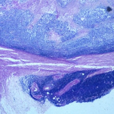
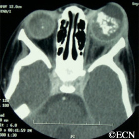
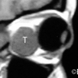
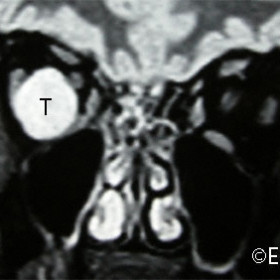
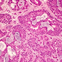
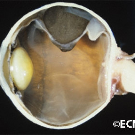
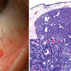
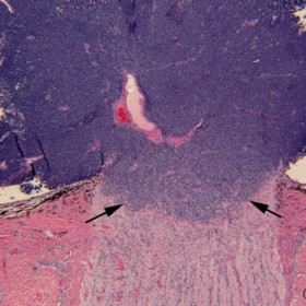
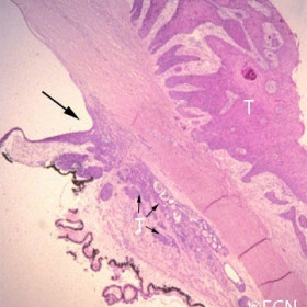
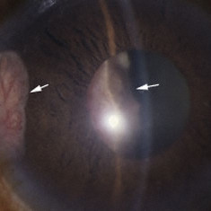
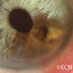
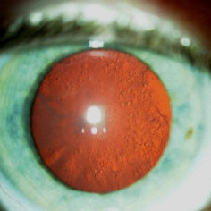
Irregularly shaped uveal melanoma
Transillumination shadow from a irregularly shaped uveal melanoma