By Paul T. Finger, MD
Case 1: Dynamic Scanning


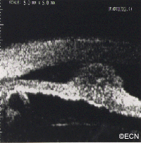
Notice the low reflective mass in the iris. There is thinning of the iris pigment epithelium and a length of relatively normal appearing iris between the posterior margin of the tumor and the ciliary body. In this case, the tumor was documented to grow and cataract surgery was contemplated. In part, due to these dynamic high-frequency ultrasound findings, excision was planned prior to dilation for cataract surgery.
This case illustrates that high-frequency ultrasound allowed for unique views of the posterior aspect of the tumor, as well as an assessment of its invasion within the iris stroma.
Case 2: A Sigh of Relief


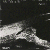
The iris cysts are the most common iris tumor sent for high-frequency ultrasonographic evaluation. Its clinical presentation is similar to seeing a localized “bulge” in the iris. Ultrasonography clearly demonstrates the cystic nature of the tumor and allows for an assessment of the adjacent ciliary body (for tumor).
Cases 3 & 4: A Unique tool


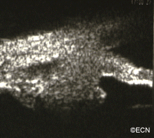
This melanoma has grown through the iris pigment (image 1) epithelium onto the anterior capsular surface. High frequency ultrasonography allows for an assessment of tumor penetration of the iris and ciliary body.
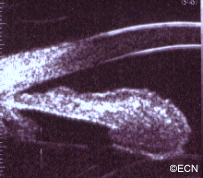
This small ciliary body melanoma (image 2) would likely have gone undetected prior to high frequency ultrasonography. Like other malignant tumors, early detection and treatment of ciliary body melanomas should improve survival.
References
- Reminick LR, Finger PT, Ritch R, Weiss S, Ishikawa H. Ultrasound biomicroscopy in the diagnosis and management of anterior segment tumors. Journal of the American Optometric Association 69(5);575-582, 1998.
- Katz NR, Finger PT, McCormick SA, Tello C, Ritch R, Sirota M, Kranz O. Ultrasound biomicroscopy in the management of malignant melanoma of the iris. The Archives of Ophthalmology 1995;113:1462-1463.


