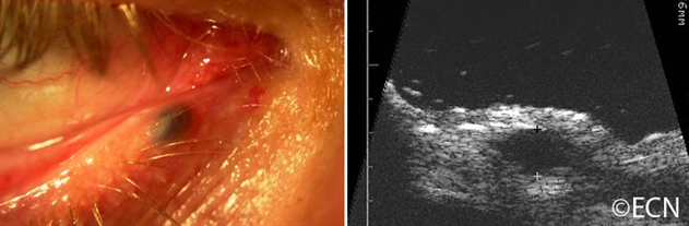
Eyelid Hydrocystoma – Left, a slit lamp photograph demonstrates a pigmented hydrocystoma (simulating a melanoma). Right, a 20 MHz high frequency ultrasound demonstrates that it is cystic.
Eyelid Hydrocystoma – Left, a slit lamp photograph demonstrates a pigmented hydrocystoma (simulating a melanoma). Right, a 20 MHz high frequency ultrasound demonstrates that it is cystic.
"Very well treated by Dr. Finger. He explained everything I needed to know about my issue with detail and attention, putting me at ease and giving me confidence to handle this problem for the rest of my life.”
– N.N.


