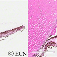
Retinoblastoma associated iris neovascularization
Retinoblastoma associated iris neovascularization (low and high power). Courtesy of Tatyana Milman, MD
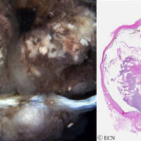
Retinoblastoma
Both gross and histopathologic images of retinoblastoma invading the optic nerve. Images courtesy of Tatyana Milman, MD
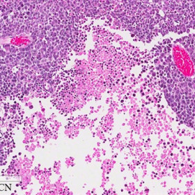
Retinoblastoma cells
Retinoblastoma cells. I high power photomicrograph courtesy of Tatyana Milman, MD
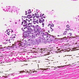
A clump of retinoblastoma cells
A clump of retinoblastoma cells located beneath the retinal pigment epithelium. Photomicrograph courtesy of Tatyana Milman, MD

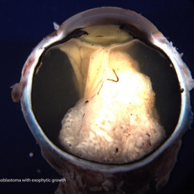
Retinoblastoma with both endophytic and exophytic
Photograph reveals a retinoblastoma with both endophytic and exophytic growth patterns (Courtesy of Tatyana Milman, MD)
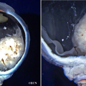
Retinoblastoma
Exophytic growth pattern, beneath total retinal detachment (Courtesy of Tatyana Millman, MD)
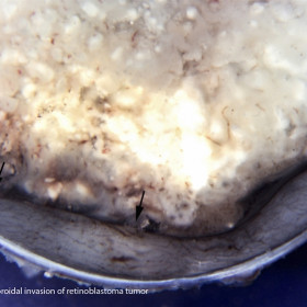
Retinoblastoma
Massive Choroidal Invasion (Courtesy of Tatyana Millman, MD)
Retinoblastoma
Stage T2b with clumps of vitreous cells courtesy of Tatyana Milman, MD
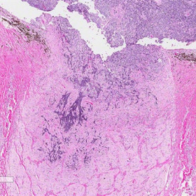
Retinoblastoma
Invasion of the optic nerve is noted. Photomicrograph courtesy of Tatyana Milman, MD


