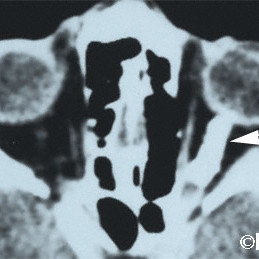
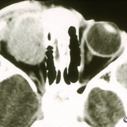
Optic nerve sheath meningioma
Optic nerve sheath meningioma (arrow head)- Computed radiographic tomography (CT).
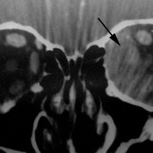
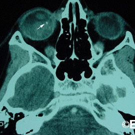
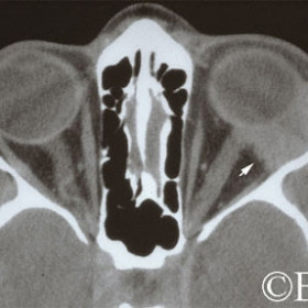
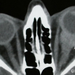
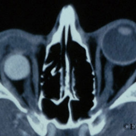
Sclerosing pseudotumor
Sclerosing pseudotumor - CT coronal image - nasal orbit- Note the irregular tumor margins.
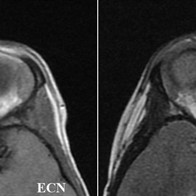
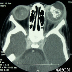
Sarcoma
Sarcoma - Soft tissue sarcoma arising from the sclera- Magnetic resonance imaging T2 - dark (see arrow)
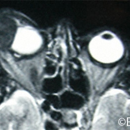
Rhabdomyosarcoma
Rhabdomyosarcoma as seen on Magnetic Resonance Imaging (MRI)
Rhabdomyosarcoma
Rhabdomyosarcoma as seen on computed radiographic tomography (CT).
Retinoblastoma
Retinoblastoma - Computed radiographic tomography demonstrates calcium (arrow) within a large intraocular retinoblastoma
Retinoblastoma
Retinoblastoma - Computed radiographic tomography (CT) demonstrates unilateral buphthalmos and bilateral intraocular calcifications.
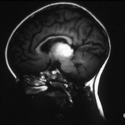
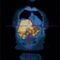
Pinealoma associated with a bilateral retinoblastoma
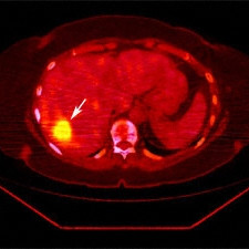
PET:CT image
PET:CT image demonstrates an isolated liver metastasis (arrow)
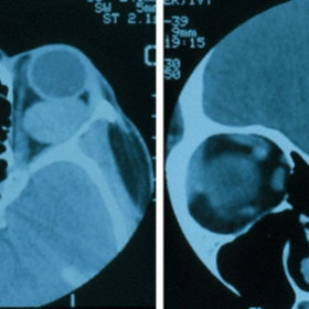
Orbital hemangioma
Orbital hemangioma - A relatively large intraconal tumor seen on axial and coronal CT images.
Adenoid cystic carcinoma
Adenoid cystic carcinoma of the lacrimal gland (arrow).
Metastatic choroidal melanoma
Metastatic choroidal melanoma - Total body PET:CT puts the form of spiral CT and the function seen with positron emission tomography on the same diagnostic page.
Malignant Lymphoma
Malignant Lymphoma involving the orbit, sclera and choroid (arrow) prior to radiation therapy
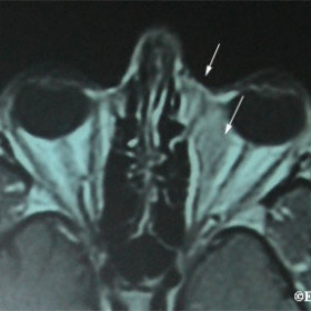
Magnetic resonance imaging
Magnetic resonance imaging (T2), demonstrates an nasal orbital lymphoma with subcutaneous extension (arrows).
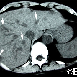
Liver metastasis
Liver metastasis - Computed axial tomography CT reveal multiple hypodense tumors (arrows).
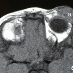
Infantile hemangioma
Infantile hemangioma - MRI image of tumor in the supero-temporal orbit (H).
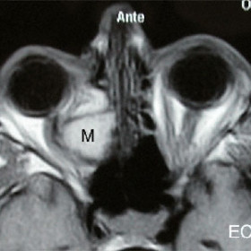
Ethmoidal mucocele
Ethmoidal mucocele - Magnetic resonance imaging (MRI) demonstrates an ethmoidal mucocele (M) displacing the optic nerve.
Enucleation Implant
Enucleation Implant - Computed radiographic tomography reveals the hyperdense (PMMA) spherical implant within the muscle cone of the left orbit.
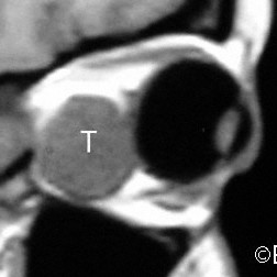
Choroidal Melanoma
MRI SAGITTAL SECTION demonstrates massive extrascleral tumor extension (T).
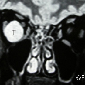
Choroidal Melanoma
Choroidal Melanoma - MRI CORONAL section demonstrates massive extrascleral tumor extension (T).


