Adenocarcinoma of the retinal pigment epithelium
Adenocarcinoma of the retinal pigment epithelium wth focal invasion of the choroid.
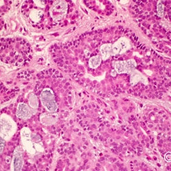
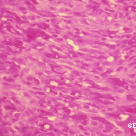
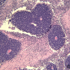
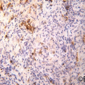
Adenoid cystic carcinoma
Capillary hemangioma specimen from a child.
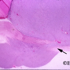
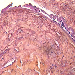
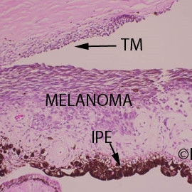
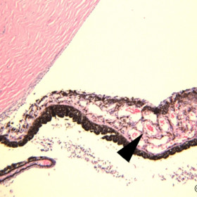
Choroidal melanoma
Choroidal melanoma - Histopathology reveals the sclerostomy (arrow) through which the retrobulbar melanoma was formed.
Choroidal Melanoma
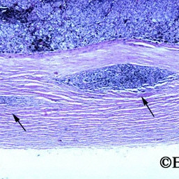
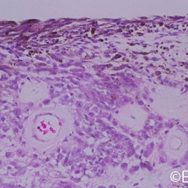
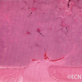
Choroidal Melanoma - As many as 50% of choroidal melanomas are thought to invade the sclera (arrows).
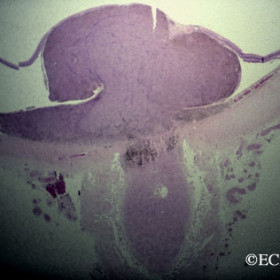
Choroidal melanoma of the optic nerve
Choroidal melanoma of the optic nerve - This circumpapillary melanoma has grown as to cover and extend into the optic nerve.
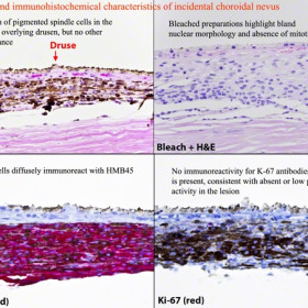
Choroidal Nevus Histopathology
Choroidal Nevus Histopathology - Courtesy of Tatyana Milman, MD
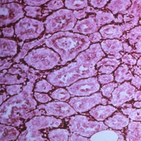
Epibulbar Oncocytoma
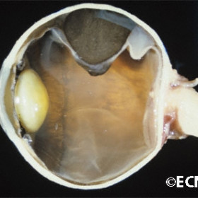
Gross pathologic specimen
Gross pathologic specimen - Section of an eye with a choroidal melanoma.
Iris Melanocytoma
Iris Melanocytoma - bleached iridectomy specimen
Iris Melanoma
Iris Melanoma - HIstopathology reveals full thickness invasion of the iris stroma by melanoma cells.
Iris Melanoma
Iris Melanoma - Diffuse melanoma with invasion of the iris stroma and trabecular meshwork.
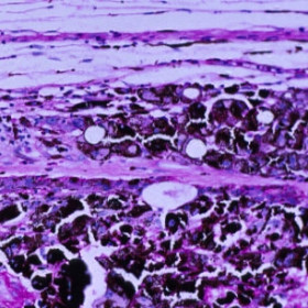
Kaposis sarcoma histology
Kaposis sarcoma histology - The tumor is almost entirely composed of blood vessels.
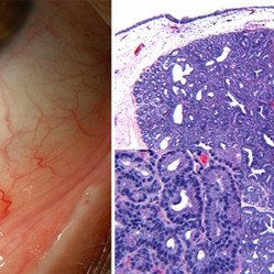
Lacrimal Gland Choristoma
Lacrimal Gland Choristoma - Histopathology images courtesy of Tatyana Milman, MD.
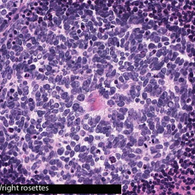
Pathology Slides
Pathology Slides - Retinoblastoma- Homer-Wright rosettes II (Courtesy of Tatyana Milman, MD)
Patient with known metastatic breast cancer presents with bilateral conjunctival metastases.
Retinoblastoma
Retinoblastoma - Tumor induced iris neovascularization stands as evidence of secondary neovascular glaucoma (Courtesy of Tatyana Millman, MD)
Retinoblastoma
Homer Wright Rosettes (arrows). Pseudorosettes formed by the arrangement of tumor cells around an area of fibrillarity, evidence of neuroblastic differentiation in a retinoblastoma (Courtesy of Tayana Millman, MD).
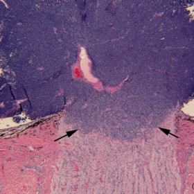
Retinoblastoma
Retinoblastoma - High power histopathology reveals tumor invasion (arrows) into the optic nerve (Courtesy of Tatyana Millman, MD)
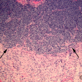
Retinoblastoma
Retinoblastoma - low power histopathology reveals tumor invasion (arrows) into the optic nerve (Courtesy of Tatyana Millman, MD)
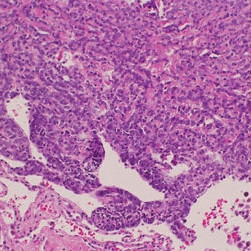
Retinoblastoma
Retinoblastoma- This retinoblastoma seed is seated below the retinal pigment epithelium, but does not extend into the subjacent choroid (Courtesy of Tatyana Millman, MD)
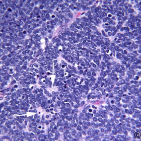
Retinoblastoma
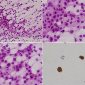
Retinoblastoma- This slide demonstrates an area of relatively undifferentiated clumps of retinoblastoma cells (Courtesy of Tatyana Millman, MD)
Retinoblastoma with intratumoral calcifications (arrows).
Ring Melanoma
Hematoxylin and eosin composite demonstrates malignant melanoma in the ciliary body and on the iris surface (Courtesy of Tatyana Millman, MD)
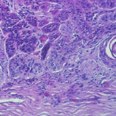
Sarcoidosis
Sarcoidosis - Iris tumor- iridectomy specimen
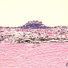
Squamous carcinoma of the conjunctiva
Squamous carcinoma of the conjunctiva - Note the tumor is invading the cornea and sclera.
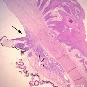
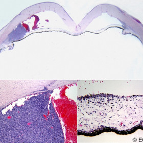
Squamous carcinoma of the conjunctiva
Squamous carcinoma of the conjunctiva (T) with intraocular invasion (arrows)
Wilm`s Tumor


