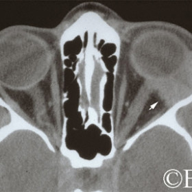
Adenoid cystic carcinoma of the lacrimal gland
Adenoid cystic carcinoma of the lacrimal gland (arrow).
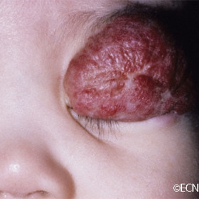
Capillary hemangioma of childhood
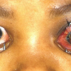
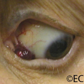
orbital extension of retinoblastoma
Clinical photograph of a child with orbital extension of retinoblastoma.
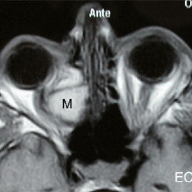
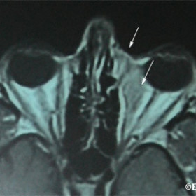
Ethmoidal mucocele (M) displacing the optic nerve
Magnetic resonance imaging (MRI) demonstrates an ethmoidal mucocele (M) displacing the optic nerve.
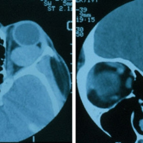
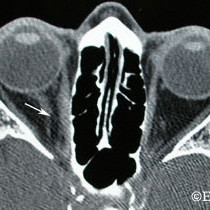
Intraconal orbital hemangioma
Intraconal orbital hemangioma - A relatively large tumor seen on axial and coronal CT images.
Nasal orbital lymphoma with subcutaneous extension
Magnetic resonance imaging (T2), demonstrates an nasal orbital lymphoma with subcutaneous extension (arrows).
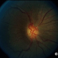
Optociliary shunt vessels
Optociliary shunt vessels induced by an optic nerve sheath meningioma.
Orbital lymphangioma
Orbital lymphangioma that invades the upper lid and has subconjunctival lymphangiectasias
Orbital lymphoma
Orbital lymphoma 6 months after external beam radiation therapy (arrow)
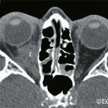
Orbital lymphoma before radiation therapy (T)

Orbital Sarcoid
Orbital Sarcoid - primarily involving the lacrimal gland
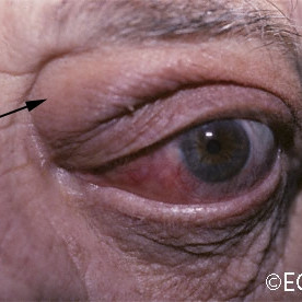
Sclerosing pseudotumor
Sclerosing pseudotumor - Note the periorbital edema (arrow) and epibulbar infiltration.
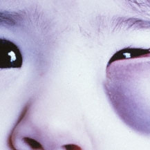
Subcutaneous capillary hemangioma
Subcutaneous capillary hemangioma can cause bluish discoloration of the eyelid


