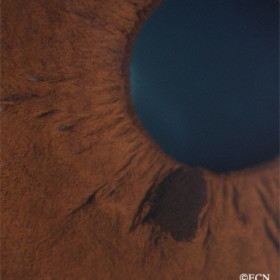
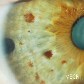
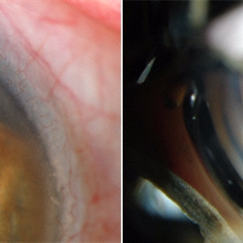
Multiple iris nevi
Multiple iris nevi on gonioscopy

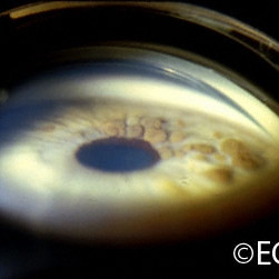
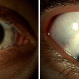
Iris neovascularization
Iris neovascularization secondary to a medium-sized choroidal melanoma with a long-standing exudative retinal detachment (prior to treatment).
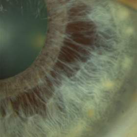
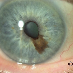
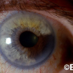
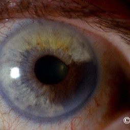
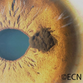
Iris Nevus
Iris Nevus - Slit lamp photography documents the appearance and distribution of this rather benign appearing iris nevus.
Iris Nevus
Iris Nevus - Note the lack of ectropion uveae, correctopia, intrinsic vascularity, or pigment dispersion.
Iris pigment epithelium (IPE) Cyst
Iris Stromal Atrophy
Iris stromal cyst
Iris stromal cyst as seen on gonioscopy
Iris Varix
Juvenile Xanthogranuloma
Juvenile Xanthogranuloma in a 2 year old male. This case had no systemic disease and the lesion resolved with steroid therapy.
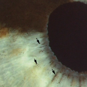
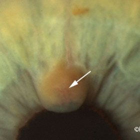
Iris Melanoma
Iris Melanoma with pigment liberation (arrow) onto the surface of the iris stroma.
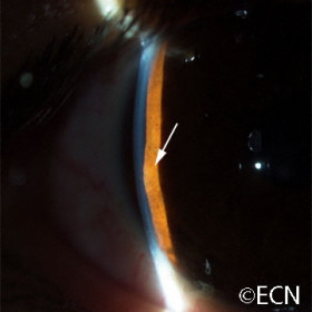
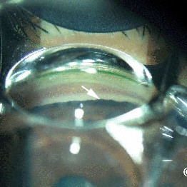
Neuro-epithelial iris cyst
Neuro-epithelial iris cyst typically presents as a bulge in the iris stroma.
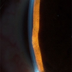
Neuroepithelial iris cyst
Neuroepithelial iris cyst - Typically presents as a focal protrusion of the iris stroma anterior to the cyst.
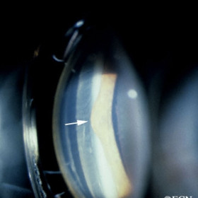
Neuroepithelial iris cyst
Neuroepithelial iris cyst - Gonioscopic image clearly demonstrates focal anterior displacement of the iris stroma (arrow).
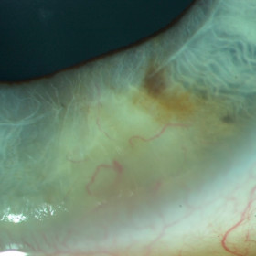
Ocular Melanosis
Ocular Melanosis - Slit lamp photography demonstrates episcleral and uveal pigmentation associated with ocular melanosis.
Pearl cyst of the iris
Pearl cyst of the iris - Epithelial implantation cyst.
Small iris melanocytoma
Small iris melanocytoma - note no evidence of ectropion, intrinsic vascularity, or sector cataract
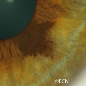
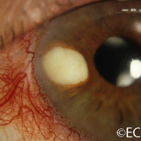
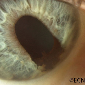
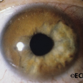
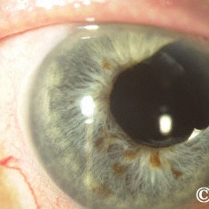
Suspicious Iris Nevus
Suspicious Iris Nevus - Note the ectropion uveae, correctopia and the variable brown and "tapioca" coloration.
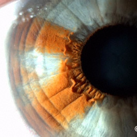
Iris Flocculi
Iris Flocculi - Hi power slit lamp photograph reveals multiple pigmented flocculi at the pupillary margin.
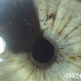
Gonioscopic appearance
Gonioscopic appearance of pigment deposition in the anterior chamber angle (arrow)
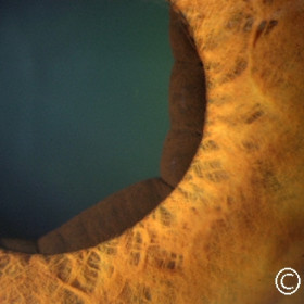
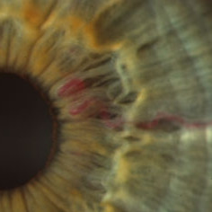
Intrinsic vascularity
Intrinsic vascularity (arrow) in a suspicous iris nevus

Intrinsic vascularity
Intrinsic vascularity seen in a suspicious iris nevus
IPE cyst
IPE cyst that eroded through the iris stroma
Iridociliary Melanoma
Iridociliary Melanoma - Note the correctopia
Iridociliary melanoma
Iridociliary melanoma - In this case the tumor has filled one sector of the anterior chamber and touches the corneal endothelium.
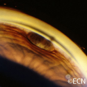
Iris Cyst
Iris Cyst - Transstromal Iris Cyst with compression of the iris stroma anterior to the tumor (arrow). High frequency ultrasound shows echolucency within the tumor.
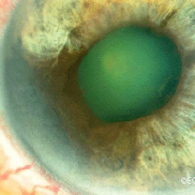
Iris ectropion uveae
Iris ectropion uveae - Induced by an iridociliary melanoma.
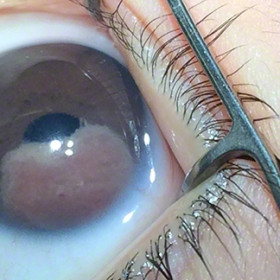
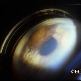
Diffuse iris melanoma
Diffuse iris melanoma - This eye was lost to secondary glaucoma.
Iris Melanocytoma
Iris Melanocytoma - Note its cobblestone surface and slight correctopia.

Iris Melanoma
Iris Melanoma - This tumor was characterized by pigment dispersion on to the iris stroma and correctopia.
Iris Melanoma
Iris Melanoma
Iris Melanoma - Gonioscopy reveals the pigmented iris tumor arising from the iris stroma. It is seen to extend from the ciliary body band into the pupil.
Iris Melanoma
Iris Melanoma - A surgical iridectomy completely removes the tumor but leaves an enlarged pupillary aperture.
Iris melanoma
Iris melanoma - Note the pigment liberated from the tumor onto the adjacent iris stroma.
Iris Melanoma
Iris Melanoma fills the angle and causes correctopia


