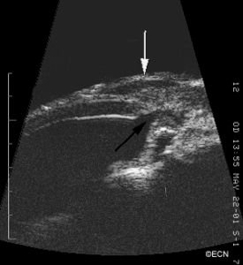
High-frequency Ultrasonography reveals the open sclerostomy (black arrow) and the overlying low-reflective tumor (white arrow). Note that the ciliary body is highly reflective (unlike the tumor). A multicystic bleb is noted on transverse section. High-frequency ultrasound obtained with the 20 MHz probe.


