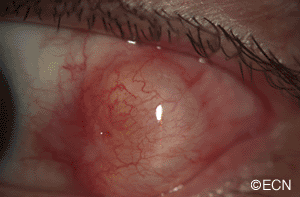
A digital slit-lamp photograph of a conjunctival cyst overlying the insertion of the lateral rectus muscle. The anterior, superior and inferior margins are clearly visible. The posterior and episcleral margins are not visible. There is also a question as to the involvement of the lateral rectus muscle.


